Laser Phase Plate Breakthrough Illuminates the Smallest Proteins in Human Cells
A revolutionary laser enhancement for cryo-electron microscopes allows scientists to clearly image the 90% of human proteins previously too small to see, unlocking a new era for structural biology and drug discovery.
By Factlen Editorial Team
- Structural Biologists
- Researchers focused on mapping the physical architecture of molecules to understand how they function.
- Biomedical Engineers
- Physicists and engineers focused on overcoming the technical limitations of scientific hardware.
- Pharmaceutical Researchers
- Industry scientists looking for new, high-resolution drug targets to cure complex diseases.
- Synthesis & Analysis
- Editorial synthesis providing broader context on the historical and medical stakes of the breakthrough.
What's not represented
- · Computational Biologists (AI structure prediction)
- · University Funding Boards
- · Clinical Trial Designers
Why this matters
For decades, scientists have been trying to design drugs for diseases while blind to the exact shape of the proteins causing them. By making the smallest, most elusive proteins visible at the atomic level, this technology directly accelerates the creation of highly targeted treatments for cancer, Alzheimer's, and autoimmune disorders.
Key points
- Cryo-electron microscopy (cryo-EM) has historically struggled to image small proteins, leaving 90% of the human proteome out of focus.
- Researchers at UC Berkeley and the Chan Zuckerberg Biohub have successfully built a 'laser phase plate' to solve this physical limitation.
- The device uses a continuous-wave laser 100 million times brighter than the sun to shift the phase of the microscope's electron beam.
- This creates massive natural contrast without blurring the image, successfully resolving small benchmark proteins like hemoglobin.
- The breakthrough paves the way for mapping intact cells and designing new targeted drugs for previously un-targetable diseases.
The human cell is a bustling, densely packed metropolis of molecular machinery, but for decades, scientists have been trying to map its intricate architecture while effectively wearing a blindfold. Cryo-electron microscopy (cryo-EM) revolutionized the field of structural biology by allowing researchers to flash-freeze proteins in vitreous ice and photograph them at the atomic level, an achievement so profound it earned the Nobel Prize in Chemistry in 2017. Yet, despite its transformative impact on our understanding of biology, cryo-EM has always harbored a massive, frustrating blind spot. Because the technique relies on firing electrons at biological samples, and because those samples are composed of light elements that are largely transparent to electrons, more than 90% of the proteins inside human cells are simply too small to generate enough contrast to be captured clearly. They remain hidden in the static.[4][5]
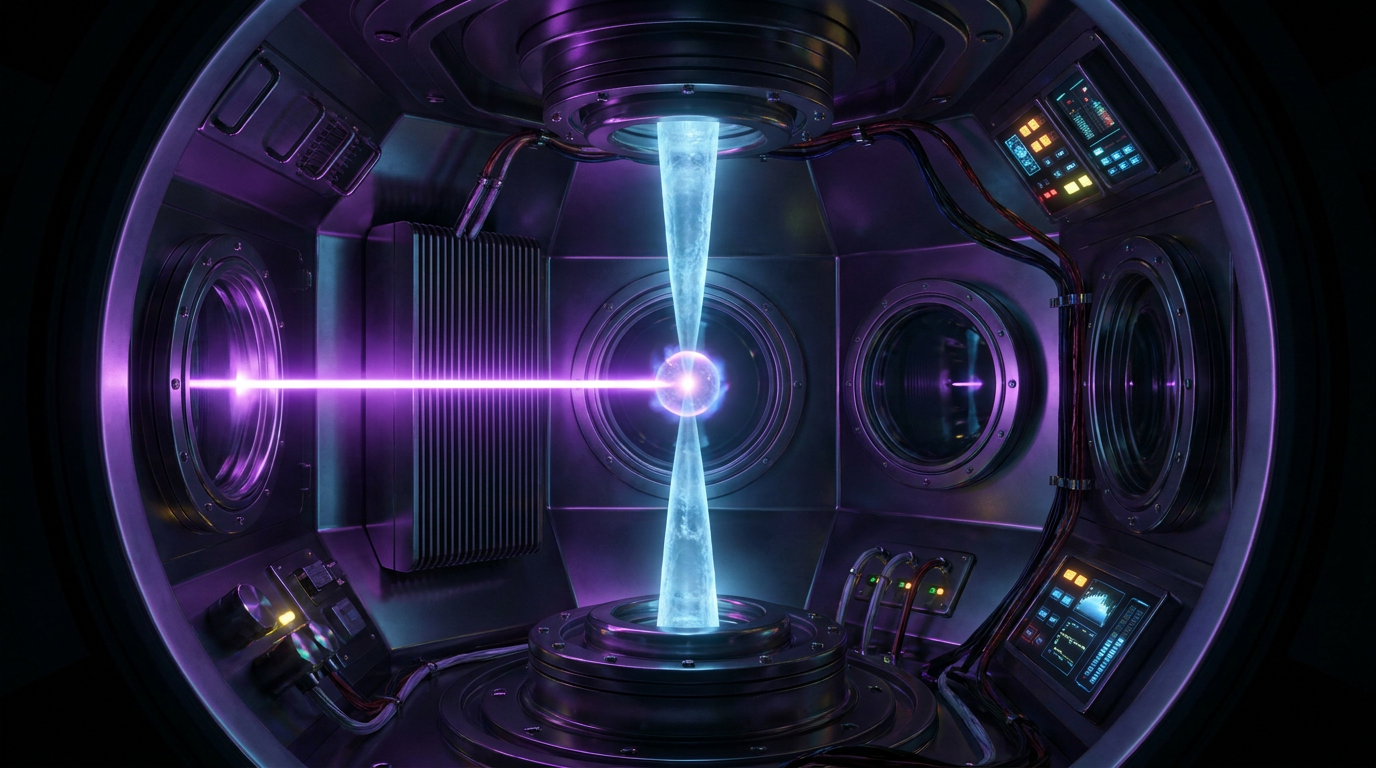
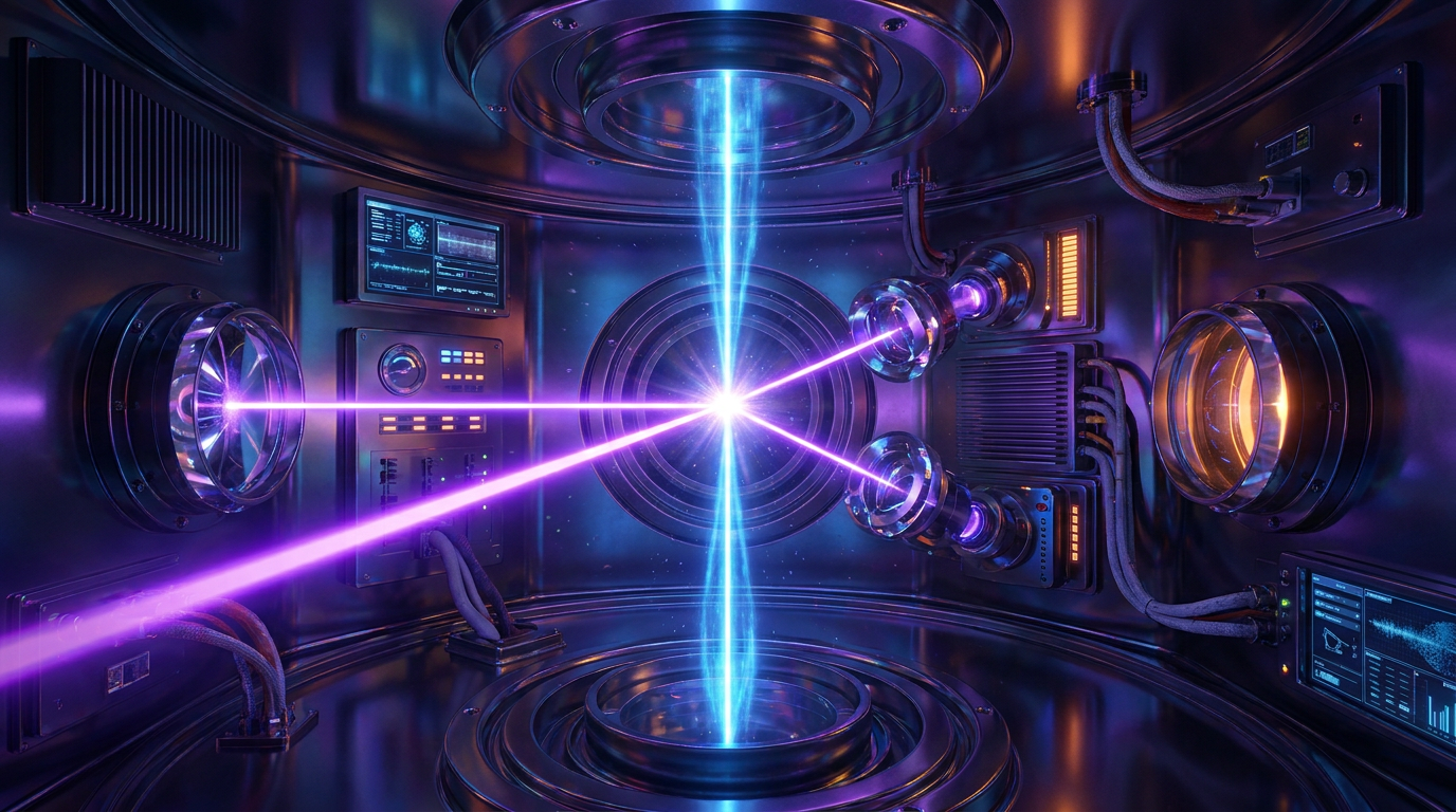
That fundamental blind spot is now being brilliantly illuminated by a feat of optical engineering that many in the physics community once considered impossible. In a landmark achievement published this week in the journal Science, a collaborative team of researchers from the University of California, Berkeley and the Chan Zuckerberg Biohub have successfully demonstrated a working 'laser phase plate'—a novel device that dramatically and cleanly boosts the contrast of cryo-EM images. By precisely crossing the microscope's delicate electron beam with a continuous-wave laser that is 100 million times brighter than the surface of the sun, the team has managed to make the invisible visible. This breakthrough effectively removes the physical limitations that have held back structural biology, opening up an entirely new view of human biology at its most fundamental level.[1][2][3]
To truly understand why this development is such a triumph, one must first understand the quantum physics of seeing the microscopic world. Biological molecules, such as proteins and nucleic acids, are primarily composed of light elements like carbon, nitrogen, and oxygen. When a high-energy electron beam passes through these biological samples in a standard cryo-EM setup, the electrons do not bounce off the atoms like light bouncing off a mirror. Instead, they pass right through the sample, merely slowing down slightly as they interact with the atomic nuclei. This interaction creates what physicists call a 'phase shift' in the electron wave, but it produces almost no visible amplitude contrast in the resulting image. It is akin to trying to see a perfectly clear pane of glass submerged in a pool of water—the information is there, but the eye cannot perceive it.[5]

For years, microscopists have solved this transparency problem by intentionally throwing the microscope's image slightly out of focus. This technique, known as 'defocusing,' creates artificial contrast by allowing the scattered and unscattered electron waves to interfere with each other, allowing the larger protein structures to finally emerge from the background noise. But defocusing comes at a steep and unavoidable cost: it inherently blurs the finest, highest-resolution details of the image. For massive protein complexes or large viruses, this trade-off is entirely acceptable, as their sheer size compensates for the blur. However, for small proteins, the defocusing blur swallows the molecule entirely, rendering it an indistinguishable, blurry smudge that is useless for scientific analysis or drug design.[1][6]
The theoretical solution to this contrast problem was actually proposed nearly a century ago for traditional light microscopes by the Dutch physicist Frits Zernike, who won a Nobel Prize in 1953 for inventing the phase-contrast microscope. Zernike realized that if you could artificially shift the phase of the unscattered light by exactly 90 degrees relative to the scattered light, the two waves would perfectly interfere with each other at the detector. This 90-degree shift converts transparent phase differences directly into stark, high-contrast visible amplitude differences without ever needing to defocus the lens. The result is a perfectly sharp, high-contrast image that retains all of its high-resolution data, a principle that revolutionized cellular biology in the 20th century.[2][6]
Applying Zernike's elegant principle to the harsh environment of electron microscopes, however, proved to be maddeningly difficult for generations of engineers. Previous attempts to build a physical phase plate for cryo-EM involved inserting ultra-thin carbon films directly into the path of the electron beam. While these carbon films successfully shifted the phase of the electrons, they suffered from a fatal flaw: the intense electron beam inevitably charged the carbon, creating localized electrostatic fields. These unpredictable electric fields perturbed the passing electron waves, entirely scrambling the resulting image and rendering the data useless. The field desperately needed a phase plate that could shift the electrons without holding a static charge, a seemingly paradoxical requirement.[6]
More than 15 years ago, UC Berkeley physicist Holger Müller and biophysicist Robert Glaeser proposed a radical, outside-the-box alternative: what if the phase plate wasn't made of physical matter at all, but of pure, concentrated light? They theorized that an intense, continuous-wave laser could generate a ponderomotive potential—a localized electromagnetic field strong enough to retard the passing electrons by exactly 90 degrees. Because the laser is made of photons, it has no physical mass and cannot accumulate an electrostatic charge, neatly bypassing the fatal flaw of the carbon films. The laser would act as an invisible, frictionless optical lens, perfectly shifting the electron beam without ever physically touching it or degrading the sample.[2][6]

Because the laser is made of photons, it has no physical mass and cannot accumulate an electrostatic charge, neatly bypassing the fatal flaw of the carbon films.
At the time of its proposal, the idea was widely dismissed by the scientific community as an engineering impossibility. Generating a continuous-wave laser of that extreme intensity inside the high-vacuum environment of an electron microscope required bouncing a beam back and forth between perfectly aligned, highly polished mirrors, creating a standing wave of staggering optical power. The engineering tolerances required for this optical cavity were so tight that even the slightest ambient vibration, acoustic noise, or microscopic thermal expansion would instantly collapse the system. Building it required marrying the bleeding-edge precision of quantum optics with the bulky, industrial reality of a commercial electron microscope.[3]
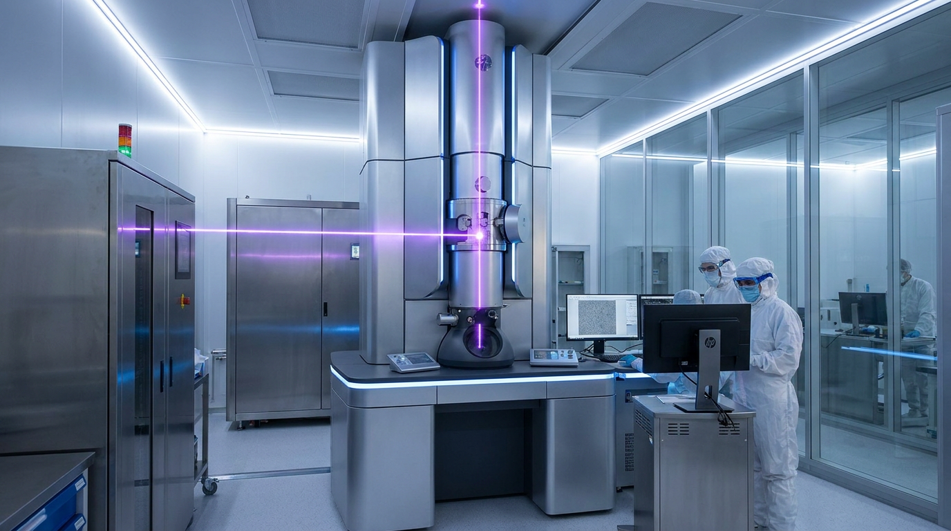
Backed by a massive, long-term financial bet from the Chan Zuckerberg Biohub, Müller's team spent the next decade methodically turning the impossible into reality. They acquired a state-of-the-art Thermo Scientific Krios cryo-electron microscope and heavily customized it, integrating a bespoke optical cavity directly into the electron column. The resulting laser phase plate generates a steady-state laser field that is one of the brightest of its kind in the world, perfectly intersecting the electron beam in a space no wider than a human hair. It represents an extraordinary convergence of precision engineering, advanced laser optics, and structural biology.[3][7]
The results of this decade-long engineering marathon, published this week, are nothing short of spectacular. To definitively prove the system's efficacy, the research team chose to image hemoglobin—the vital, oxygen-carrying protein found in red blood cells. Hemoglobin is notoriously difficult for conventional cryo-EM to capture because its small size places it right at the absolute lower limit of what current machines can resolve. It is widely considered the ultimate benchmark test for any new advancement in electron microscopy, separating theoretical improvements from genuine, practical breakthroughs.[1][2]
With the laser phase plate engaged, the hemoglobin molecules snapped into brilliant, high-contrast focus without the need for any image-degrading defocusing. The team demonstrated that the smaller the biological sample, the more profound the improvement in resolution and contrast provided by the laser. The resulting 3D reconstructions revealed the atomic architecture of the protein with unprecedented clarity. For structural biologists who have spent their careers squinting at blurry, low-contrast data, it was the equivalent of being handed a pair of high-prescription glasses after years of near-blindness.[1][7]

The implications of this breakthrough for modern medicine and pharmaceutical development are profound and immediate. The vast majority of modern pharmaceuticals are developed using a technique called structure-based drug design. In this process, scientists map the exact, three-dimensional atomic shape of a disease-causing protein, and then computationally design a chemical 'key' that perfectly fits into the protein's specific 'lock' to disable it. If you cannot see the lock, you cannot design the key, which is why high-resolution structural data is the lifeblood of the pharmaceutical industry.[4][5]
Because conventional cryo-EM could only clearly resolve the largest 10% of human proteins, the vast majority of potential drug targets remained locked in a blurry, inaccessible void. The laser phase plate cracks this void wide open. By providing the contrast necessary to image small molecules, this technology potentially allows researchers to map the atomic structures of the remaining 90% of the human proteome. This includes countless elusive proteins that are deeply implicated in aggressive cancers, neurodegenerative diseases like Alzheimer's, and complex autoimmune disorders, transforming them from mysteries into viable therapeutic targets.[3][7]
But the ambition of the research team extends far beyond imaging isolated, purified proteins in a vacuum. The next great frontier in structural biology is cryo-electron tomography (cryo-ET), an advanced technique that involves imaging intact, frozen cells to see exactly how proteins interact with their neighbors in their native, crowded environments. Cryo-ET suffers from even worse contrast issues than standard cryo-EM because the electron beam must penetrate the entire dense thickness of the cellular environment, resulting in incredibly noisy, low-contrast data.[2][3]

To conquer this final frontier, the Chan Zuckerberg Biohub is already actively testing a next-generation, dual-laser phase plate specifically designed to supercharge cryo-ET. By applying this massive, laser-driven contrast boost to whole cells, scientists hope to eventually generate comprehensive, atomic-level 3D maps of the entire cellular interior. This would allow researchers to watch the molecular machinery of life operate in its natural context, observing exactly how viruses hijack cellular factories or how genetic mutations physically manifest as disease within the cell.[3][5]
"It's like seeing first light through a telescope," noted Bridget Carragher, the founding technical director of imaging at the Biohub, capturing the magnitude of the moment. The laser phase plate is not just an incremental hardware upgrade; it is a fundamental paradigm shift in the history of microscopy. After 15 years of relentless engineering against overwhelming odds, the lights have finally been turned on inside the cell, and the era of comprehensive, atomic-level structural biology has officially begun.[3][5]
How we got here
1953
Frits Zernike wins the Nobel Prize in Physics for inventing the phase-contrast light microscope, proving that phase shifts can create contrast.
2010s
Advances in direct electron detectors trigger the 'resolution revolution' in cryo-EM, making it a dominant tool in structural biology.
2017
The Nobel Prize in Chemistry is awarded to Jacques Dubochet, Joachim Frank, and Richard Henderson for developing cryo-electron microscopy.
2021
The Chan Zuckerberg Biohub makes a major funding bet on UC Berkeley's Holger Müller to build a working laser phase plate.
June 2026
Researchers successfully demonstrate the laser phase plate in a state-of-the-art microscope, capturing high-contrast images of small proteins like hemoglobin.
Viewpoints in depth
Structural Biologists
Researchers focused on mapping the physical architecture of molecules to understand how they function.
For decades, structural biologists have been frustrated by the physical limits of electron scattering. They view the laser phase plate as the holy grail of their field, as it eliminates the need to defocus images—a compromise that previously destroyed high-resolution data for small molecules. Their immediate focus is deploying this tool to map the 90% of the human proteome that has historically been invisible to cryo-EM, opening up entirely new avenues of basic biological research.
Biomedical Engineers
Physicists and engineers focused on overcoming the technical limitations of scientific hardware.
Engineers and physicists view this breakthrough as a triumph over seemingly impossible technical constraints. Generating a continuous-wave laser 100 million times brighter than the sun inside the delicate vacuum of an electron microscope required unprecedented precision and stability. They are already looking ahead to the next iteration: dual-laser systems designed to penetrate entire intact cells for cryo-electron tomography, pushing the boundaries of optical physics even further.
Pharmaceutical Researchers
Industry scientists looking for new, high-resolution drug targets to cure complex diseases.
The pharmaceutical industry relies heavily on structure-based drug design, which requires an exact 3D model of a target protein to develop a chemical inhibitor. Because conventional cryo-EM could only resolve massive protein complexes, many disease pathways remained un-targetable. Researchers in this camp see the laser phase plate as a massive catalyst for discovering new drugs for cancer, Alzheimer's, and autoimmune disorders, as it finally allows them to see the 'locks' they need to build 'keys' for.
What we don't know
- How quickly this highly complex, bespoke laser technology can be commercialized and scaled to research laboratories worldwide.
- Whether the dual-laser phase plate currently in development will successfully penetrate the thickness of whole mammalian cells for cryo-electron tomography.
- The exact degree to which AI protein-folding models (like AlphaFold) will complement or compete with these newly acquired physical structures.
Key terms
- Cryo-electron microscopy (cryo-EM)
- A technique that uses an electron beam to photograph rapidly frozen biological molecules at the atomic level.
- Phase shift
- A change in the timing or alignment of a wave (like light or electrons) as it passes through a material, which can be used to create visual contrast.
- Defocusing
- The practice of intentionally blurring a microscope image to artificially increase the contrast of transparent objects.
- Proteome
- The entire set of proteins expressed by a genome, cell, tissue, or organism at a certain time.
- Cryo-electron tomography (cryo-ET)
- An advanced imaging technique that captures multiple 2D images of an intact cell from different angles to build a 3D model of its interior.
Frequently asked
What is cryo-electron microscopy (cryo-EM)?
It is a Nobel Prize-winning technique that fires a beam of electrons at rapidly frozen biological samples to capture atomic-level 3D images of proteins and viruses.
Why couldn't cryo-EM see small proteins before?
Biological molecules are mostly transparent to electrons. To create contrast, scientists had to blur the image slightly (defocusing), which washed out the fine details of small proteins.
How does the laser phase plate fix this problem?
It uses an intensely bright laser to shift the phase of the electron beam by 90 degrees. This creates massive natural contrast without needing to blur the image, keeping small details perfectly sharp.
Why is seeing small proteins so important?
Over 90% of the proteins in human cells are small. Seeing their exact physical shape is the crucial first step in designing new drugs to target them when they cause disease.
Sources
Source coverage
7 outlets
4 viewpoints surfaced


[1]ScienceStructural Biologists
Laser phase plate improves structure determination of small proteins by cryo-EM
Read on Science →[2]UC BerkeleyStructural Biologists
A breakthrough in electron microscopy delivers sharper images of our body's tiniest proteins
Read on UC Berkeley →[3]Chan Zuckerberg BiohubBiomedical Engineers
Making the invisible visible: A landmark achievement in microscopy
Read on Chan Zuckerberg Biohub →[4]NatureStructural Biologists
An innovative technology boosts image quality for protein structures
Read on Nature →[5]Factlen Editorial TeamSynthesis & Analysis
Synthesis by Factlen editorial team
Read on Factlen Editorial Team →[6]National Institutes of HealthBiomedical Engineers
Laser phase plate provides stable and tunable phase shift for TEM
Read on National Institutes of Health →[7]News-MedicalPharmaceutical Researchers
Laser phase plate dramatically improves contrast of cryo-electron microscopes
Read on News-Medical →

More in science
See all 20 stories →Structural Biology
A 75-Kilowatt Laser Just Unlocked the Invisible World of Human Proteins
0 sources
Wind Power
Renewable Energy Groups Sue Pentagon Over 'De Facto Moratorium' on Wind Farms
0 sources

Cryo-EM Tech
Laser Phase Plate Breakthrough Illuminates the Invisible 90% of the Human Proteome
0 sources
Wind Power Policy
Renewable Energy Groups Sue Pentagon Over 'Total Halt' of Wind Farm Reviews
0 sources

Every angle. Every day.
Get science stories with full source coverage and perspective breakdowns delivered to your inbox.