A 75-Kilowatt Laser Just Unlocked the Invisible World of Human Proteins
A breakthrough 'laser phase plate' for electron microscopes dramatically boosts image contrast, allowing scientists to finally see the microscopic proteins that drive human health and disease.
By Factlen Editorial Team
- Structural Biologists
- Focused on achieving atomic-level resolution of purified proteins to understand their fundamental architecture.
- Cell Biologists
- Prioritize the ability to see proteins interacting in their native, crowded cellular environments via tomography.
- Microscopy Engineers
- Focused on the monumental physics challenge of stabilizing a high-power laser inside a vacuum column.
- Pharmaceutical Researchers
- Focused on the downstream applications for structure-based drug design and targeting previously invisible proteins.
What's not represented
- · Commercial microscope manufacturers
- · Computational biologists
Why this matters
More than 90% of the proteins in the human body have historically been too small to image clearly, making it incredibly difficult to design drugs that target them. This optical breakthrough brings those invisible molecules into sharp focus, promising to radically accelerate the development of treatments for everything from rare genetic disorders to cancer.
Key points
- Standard cryo-electron microscopes struggle to image proteins smaller than 70 kilodaltons due to low contrast.
- Over 90% of the human proteome falls below this size threshold, making most proteins invisible to current technology.
- A new 'laser phase plate' uses a 75-kilowatt laser to shift the electron beam's phase, massively boosting image contrast.
- The technology successfully imaged hemoglobin (64 kDa) and aims to eventually resolve proteins as small as 17 kDa.
- The breakthrough will revolutionize cryo-electron tomography, allowing scientists to see proteins operating inside intact cells.
For decades, the ultimate dream of structural biologists has been to peer inside a living cell and watch its molecular machinery in action. While modern science has successfully mapped the human genome, seeing the actual proteins that carry out those genetic instructions remains a profound physical challenge. The cell is a crowded, chaotic environment, and the proteins that drive health and disease are infinitesimally small. To see them, scientists rely on cryo-electron microscopy (cryo-EM), a Nobel Prize-winning technique that fires electrons at flash-frozen biological samples. Yet, despite its revolutionary impact on drug discovery over the last decade, cryo-EM has a critical blind spot: it struggles to generate clear images of small molecules. Because biological samples are fragile, the electron beam must be kept weak to avoid incinerating the specimen, resulting in images with notoriously low contrast.[1][2]
This contrast limitation has created a formidable "size barrier" in structural biology. Standard cryo-EM machines excel at imaging massive molecular complexes, such as whole viruses or large ribosomes, but they falter when targeting proteins smaller than roughly 70 kilodaltons. This is a staggering limitation, as approximately 90 percent of the proteins in the human proteome fall below this threshold. Most of the cellular actors responsible for disease, metabolism, and cellular signaling remain too faint and blurry to be captured clearly. To study them, researchers have historically been forced to extract these proteins from the cell, crystallize them, or study them in artificial isolation—stripping away the vital context of their native environment.[3][4]

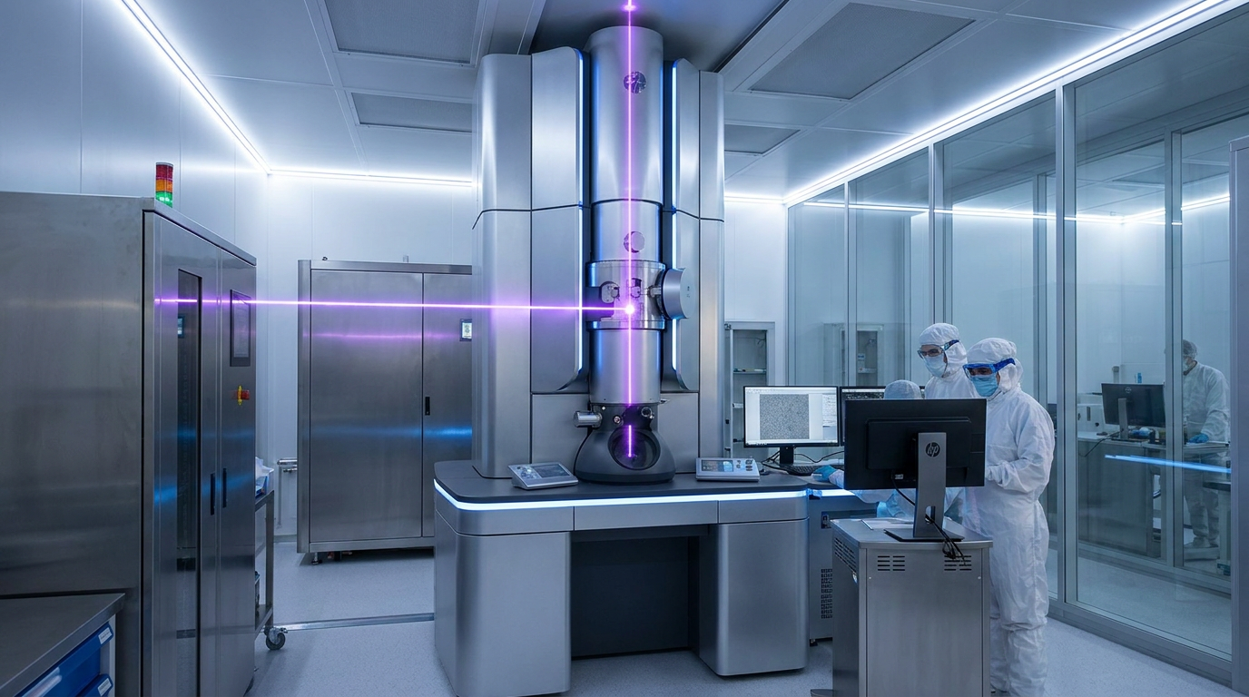
Now, a major technological leap promises to shatter this barrier. Researchers at the University of California, Berkeley, and the Chan Zuckerberg Biohub have successfully integrated a "laser phase plate" into state-of-the-art cryo-electron microscopes. Detailed in a series of recent publications, this bespoke device dramatically amplifies the contrast of electron images without requiring a destructive increase in the electron dose. By solving a physics problem that has vexed engineers for over 15 years, the team has opened an entirely new window into human biology, allowing scientists to image proteins down to 50 kilodaltons with unprecedented clarity.[2][4]
The mechanism behind the laser phase plate relies on a concept first pioneered for light microscopes nearly a century ago: phase contrast. When an electron beam passes through a biological sample, the electrons do not just scatter; their wave phase shifts slightly. However, this subtle phase shift is invisible to standard detectors. To convert it into visible contrast, the unscattered electrons must be delayed by exactly 90 degrees relative to the scattered ones. Previous attempts to achieve this in electron microscopes used physical carbon films, but these "solid" phase plates quickly accumulated electrostatic charge, ruining the image. The new system brilliantly replaces the physical film with pure light.[2][5]
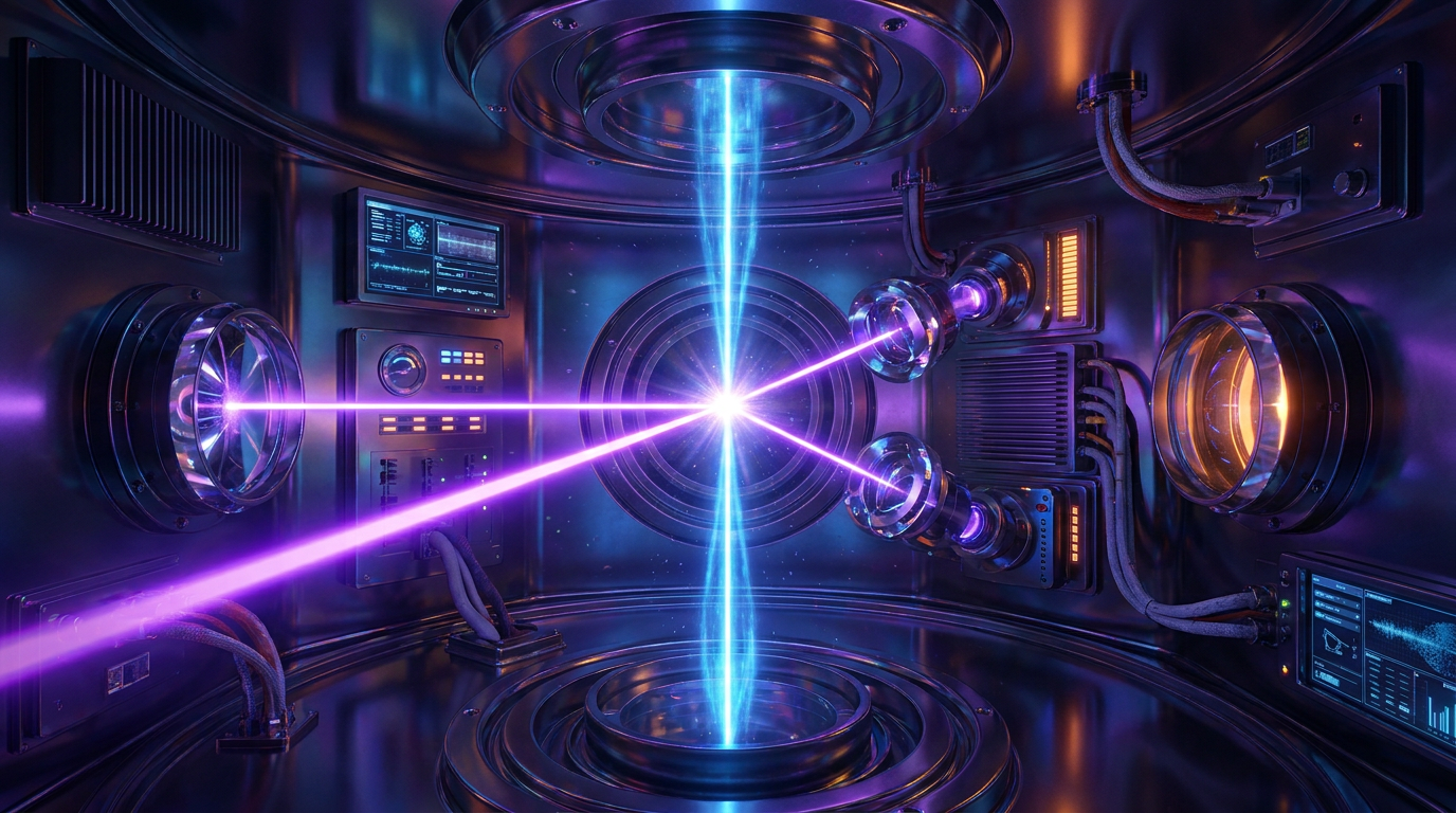
Inside the vacuum column of the electron microscope, the researchers constructed a microscopic optical cavity. They pump a continuous-wave laser into this mirrored trap, amplifying it to a staggering 75 kilowatts of power concentrated into a beam just a few microns wide. When the electron beam passes through this intense field of light, the ponderomotive potential of the laser shifts the electrons' phase by the required 90 degrees. Because the phase plate is made entirely of photons, there is no physical material to accumulate a static charge. This results in a stable, highly tunable contrast boost that leaves the fragile biological sample completely unharmed.[2][5]

Inside the vacuum column of the electron microscope, the researchers constructed a microscopic optical cavity.
The immediate impact of this contrast boost is the ability to image significantly smaller proteins. To prove the system's efficacy, the research team targeted hemoglobin, the oxygen-carrying protein in human blood. At roughly 64 kilodaltons, hemoglobin sits at the very edge of what conventional cryo-EM can resolve, typically yielding noisy, low-resolution blobs. When imaged using the laser phase plate, the contrast improved so dramatically that the team could reconstruct a high-resolution, three-dimensional model of the protein. The researchers note that the smaller the sample, the more pronounced the improvement, validating the laser's potential to unlock the vast majority of the human proteome.[3][4]
While imaging purified small proteins is a milestone, the most profound application of the laser phase plate lies in a related technique called cryo-electron tomography (cryo-ET). Unlike standard cryo-EM, which averages thousands of identical purified proteins to create a single structure, cryo-ET takes multiple angular snapshots of an intact, flash-frozen cell to build a 3D map of its interior. It allows scientists to see proteins operating in their natural, crowded habitat. However, because the electron dose must be spread across dozens of snapshots to avoid destroying the cell, the signal-to-noise ratio in cryo-ET is exceptionally poor, making it incredibly difficult to identify specific proteins within the cellular "soup."[1][3]
The laser phase plate fundamentally alters the math of cryo-ET. By massively increasing the baseline contrast of every snapshot, the technology acts like a spotlight in a dark forest, illuminating the faint outlines of small proteins against the dense background of the cell. Researchers anticipate that this will allow them to map the intricate protein-to-protein interactions that drive cellular function and viral infection directly within the cell. As one Biohub director noted, the ability to see these interactions in their native context has been the ultimate goal of cell biologists for decades, and the laser phase plate is the key to finally realizing it.[3][4]

Despite its transformative potential, the technology remains in its infancy and faces significant engineering hurdles before it can be widely adopted. Aligning a 75-kilowatt laser cavity inside the cramped, vibration-sensitive column of an electron microscope is a monumental physics challenge. The current prototypes at UC Berkeley and the CZ Biohub are highly customized, bespoke instruments that require specialized expertise to operate and maintain. The extreme precision required to keep the laser focused and stable means that commercializing the technology for standard laboratory use will require years of refinement and miniaturization.[4][6]
Furthermore, while the current system has successfully imaged proteins down to 50 kilodaltons, the theoretical limit of the technology is much lower. The engineering teams are already working on next-generation designs aimed at doubling the laser power and halving the focus size. If successful, this would optimize the phase shift for the 300-kiloelectron-volt beams preferred in advanced cryo-EM, potentially pushing the resolution limit down to 17 kilodaltons. Achieving this milestone would bring nearly every medically relevant protein into sharp focus, fundamentally accelerating the pace of structural biology.[2][5]

For the pharmaceutical industry, the implications of this enhanced visibility are profound. Modern drug design relies heavily on structure-based methods, where researchers use cryo-EM to see exactly how a small-molecule drug binds to a target protein. If the target protein is too small or flexible to be imaged clearly, designing an effective drug becomes a process of trial and error. By bringing the remaining 90 percent of the proteome into view, the laser phase plate will allow pharmaceutical researchers to rationally design targeted therapies for a vast array of previously "undruggable" diseases, from rare genetic disorders to aggressive cancers.[1][6]
The successful deployment of the laser phase plate marks a paradigm shift in how humanity observes the building blocks of life. By solving the contrast problem that has long bottlenecked electron microscopy, physicists and biologists have forged a tool that will ripple across the pharmaceutical and medical landscapes. As the technology matures and becomes more accessible, it promises to transition structural biology from studying isolated parts in a vacuum to observing the complex, dynamic machinery of the cell in its full, natural context.[4][6]
How we got here
1953
Frits Zernike is awarded the Nobel Prize in Physics for inventing phase-contrast optical microscopy.
2017
The Nobel Prize in Chemistry is awarded to the developers of cryo-electron microscopy (cryo-EM).
2019
Early theoretical frameworks and the first proof-of-concept for laser phase manipulation in electron microscopes are published.
June 2026
Researchers successfully demonstrate the laser phase plate integrated into state-of-the-art cryo-EM machines, imaging small proteins with unprecedented clarity.
Viewpoints in depth
Structural Biologists
Focused on achieving atomic-level resolution of purified proteins to understand their fundamental architecture.
For structural biologists, the primary goal is to map the exact atomic coordinates of a protein to understand how its shape dictates its function. This camp views the laser phase plate as a critical tool to expand the catalog of known protein structures. By overcoming the 70-kilodalton size barrier, they can finally apply high-resolution single-particle analysis to the vast majority of the human proteome that has thus far evaded characterization, filling in massive blanks in our understanding of molecular biology.
Cell Biologists
Prioritize the ability to see proteins interacting in their native, crowded cellular environments via tomography.
Cell biologists argue that studying a purified protein in a vacuum is like studying a single gear removed from a watch; it tells you little about how the machine actually ticks. This camp is most excited by the laser phase plate's application to cryo-electron tomography (cryo-ET). Because the laser provides such a massive boost to baseline contrast, it makes it possible to identify specific, small proteins amidst the dense, chaotic background of an intact cell, allowing researchers to map the actual interactions that drive life and disease.
Microscopy Engineers
Focused on the monumental physics challenge of stabilizing a high-power laser inside a vacuum column.
While biologists focus on the downstream applications, the engineering community views the laser phase plate as a triumph of applied physics. Aligning a 75-kilowatt laser cavity inside the vibration-sensitive, magnetically shielded column of an electron microscope was long considered nearly impossible. Engineers emphasize that while the prototypes work beautifully, the next major hurdle is miniaturizing and stabilizing the technology so that it can be manufactured at scale and operated by biologists rather than dedicated laser physicists.
What we don't know
- How quickly the bespoke laser phase plate prototypes can be commercialized and integrated into standard laboratory microscopes.
- Whether the next-generation designs will successfully reach the theoretical resolution limit of 17 kilodaltons.
- How computational pipelines will adapt to process the massive influx of high-contrast data generated by the new sensors.
Key terms
- Cryo-electron microscopy (cryo-EM)
- A technique that fires electrons at flash-frozen biological samples to determine the 3D structure of molecules at near-atomic resolution.
- Cryo-electron tomography (cryo-ET)
- An advanced imaging technique that takes multiple angular snapshots of an intact cell to build a 3D map of proteins in their natural environment.
- Phase contrast
- An optical technique that converts invisible shifts in a wave's phase (caused by passing through a transparent sample) into visible changes in brightness.
- Proteome
- The entire set of proteins that is, or can be, expressed by a genome, cell, tissue, or organism at a certain time.
- Kilodalton (kDa)
- A unit of mass used to express the molecular weight of proteins and other large biological molecules.
- Ponderomotive potential
- The physical force exerted by a highly intense, oscillating electromagnetic field (like a laser) on a charged particle (like an electron).
Frequently asked
Why couldn't scientists just use a brighter electron beam?
Biological samples are incredibly fragile. Increasing the intensity of the electron beam to get a better picture would incinerate the flash-frozen proteins before an image could be captured.
What exactly is a phase plate?
It is a device that delays the unscattered waves passing through a sample by 90 degrees. When these delayed waves recombine with the scattered waves, they create visible contrast, making transparent objects visible.
Why is the laser phase plate better than older versions?
Previous phase plates were made of physical carbon films, which quickly accumulated static electricity from the electron beam and ruined the image. The laser phase plate uses pure light, so it cannot hold a static charge.
How will this technology affect medicine?
By allowing scientists to clearly see proteins that were previously too small to image, pharmaceutical companies can rationally design new drugs that bind precisely to these targets, accelerating treatments for many diseases.
Sources
Source coverage
6 outlets
4 viewpoints surfaced


[1]NatureStructural Biologists
An innovative technology boosts image quality for protein structures
Read on Nature →[2]UC BerkeleyMicroscopy Engineers
Laser phase plate improves structure determination of small proteins by cryo-EM
Read on UC Berkeley →[3]Chan Zuckerberg BiohubCell Biologists
A Pulsed Laser Phase Plate for High-Resolution Cryo-electron Tomography
Read on Chan Zuckerberg Biohub →[4]News MedicalPharmaceutical Researchers
Laser phase plate dramatically improves contrast of cryo-EM images
Read on News Medical →[5]NIH PubMedStructural Biologists
Electron phase manipulation with a high-intensity continuous-wave laser beam
Read on NIH PubMed →[6]Factlen Editorial TeamPharmaceutical Researchers
Synthesis by Factlen editorial team
Read on Factlen Editorial Team →

More in science
See all 19 stories →Wind Power
Renewable Energy Groups Sue Pentagon Over 'De Facto Moratorium' on Wind Farms
0 sources

Cryo-EM Tech
Laser Phase Plate Breakthrough Illuminates the Invisible 90% of the Human Proteome
0 sources
Wind Power Policy
Renewable Energy Groups Sue Pentagon Over 'Total Halt' of Wind Farm Reviews
0 sources

Artemis Architecture
The Engineering Reality Behind NASA's Artemis III Restructure
0 sources
Every angle. Every day.
Get science stories with full source coverage and perspective breakdowns delivered to your inbox.