Physicists Supercharge Cryo-Electron Microscopes With Lasers to Image the Body's Tiniest Proteins
Researchers at UC Berkeley and the Chan Zuckerberg Biohub have successfully integrated a high-powered laser phase plate into a cryo-electron microscope. The engineering breakthrough dramatically boosts image contrast, allowing scientists to visualize small proteins and cellular structures that were previously invisible.
By Factlen Editorial Team
- Structural Biologists
- Focus on the ability to image smaller proteins and intact cells, which will fundamentally accelerate drug discovery and cellular mapping.
- Optical Physicists
- Focus on the technical achievement of manipulating electron beams with high-intensity lasers without causing electrostatic charging.
- Medical & Tech Analysts
- Focus on the downstream impact of mapping the 90% of the human proteome previously invisible to standard electron microscopy.
What's not represented
- · Microscope Manufacturers
- · Computational Biologists
Why this matters
By making the vast majority of human proteins visible for the first time, this breakthrough will allow scientists to see exactly how diseases operate at the atomic level. This unprecedented clarity is expected to dramatically accelerate the discovery and design of new life-saving drugs.
Key points
- Researchers successfully integrated a laser phase plate into a cryo-electron microscope.
- The technology uses a 75-kilowatt laser to shift the phase of the electron beam, dramatically boosting contrast.
- It lowers the size limit of visible proteins from 70 kilodaltons to 50 kilodaltons, with a goal of 17 kilodaltons.
- The breakthrough will allow scientists to image more than 50% of the human proteome, up from less than 10%.
- It paves the way for mapping the real-time interactions of proteins inside intact, living cells.
The human cell is a bustling metropolis of molecular machines, but for decades, scientists have been forced to study it through a frosted window. While modern imaging techniques have mapped the broad strokes of cellular architecture, the intricate, real-time interactions of the proteins that actually drive life—and disease—have remained frustratingly out of focus.[5][8]
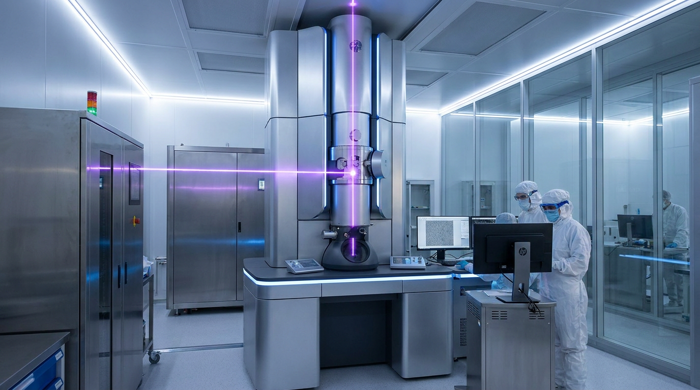
That window is finally clearing. In a landmark achievement published this week, researchers from UC Berkeley and the Chan Zuckerberg Biohub have successfully integrated a "laser phase plate" into a state-of-the-art cryo-electron microscope. This engineering feat effectively supercharges the microscope's contrast, allowing biologists to capture sharp images of molecules that were previously too small or too faint to see.[4][5]
To understand the magnitude of this breakthrough, one must look at the current gold standard for molecular imaging: cryo-electron microscopy (cryo-EM). The technique, which won the Nobel Prize in Chemistry in 2017, involves flash-freezing biological samples and bombarding them with electrons to deduce their atomic structure. It revolutionized structural biology and accelerated the development of countless drugs.[1][2]
Yet, cryo-EM has a massive blind spot. Because biological molecules are mostly made of light elements like carbon and oxygen, they barely scatter the electron beam, resulting in images with notoriously low contrast. To compensate, scientists have historically relied on studying massive protein complexes, leaving the smaller components of the cell completely invisible.[6][8]
The numbers highlight the limitation: standard cryo-EM struggles to resolve anything smaller than 70 kilodaltons. Unfortunately, more than 90 percent of the proteins in the human proteome fall below that threshold. For the vast majority of the molecular machines inside our bodies, standard cryo-EM simply cannot generate enough signal-to-noise ratio to produce a clear picture.[2][5]

The solution to this problem is rooted in a nearly century-old optical trick. In the 1930s, Dutch physicist Frits Zernike realized that when light passes through a transparent biological sample, its phase shifts slightly. By inserting a physical "phase plate" into the microscope, he converted these invisible phase shifts into stark, visible contrast—a discovery that earned him a Nobel Prize in 1953.[1][2]
For decades, physicists have tried to adapt Zernike’s phase contrast technique for electron microscopes. The concept is identical: shift the phase of the unscattered electron beam by 90 degrees relative to the scattered beam to maximize contrast. However, physical phase plates made of carbon or other materials quickly accumulate electrostatic charge when bombarded by electrons, rendering them useless.[3][6]
For decades, physicists have tried to adapt Zernike’s phase contrast technique for electron microscopes.
The UC Berkeley team, led by physicist Holger Müller, spent 15 years pursuing a radical alternative: using light to manipulate electrons. Instead of a physical plate, they engineered a system where the electron beam passes through an incredibly intense, focused laser beam.[4][5]
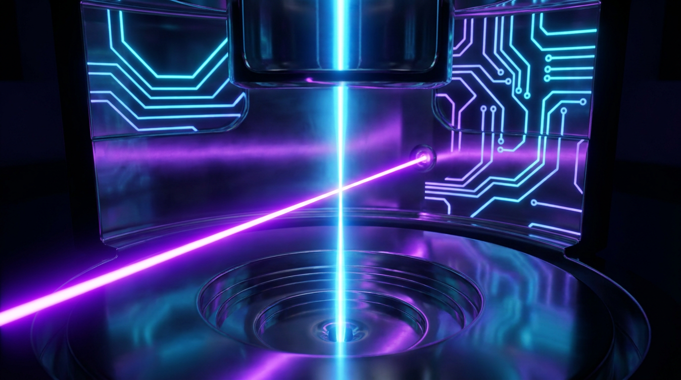
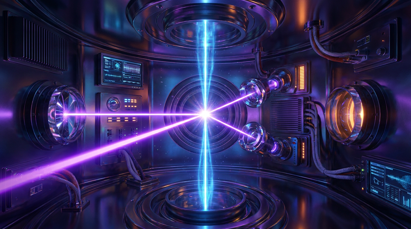
The engineering required to achieve this is staggering. The team built a mirrored cavity inside the microscope that traps and amplifies a continuous-wave laser, concentrating 75 kilowatts of power into a space just a few microns wide. This creates a "ponderomotive potential"—an electromagnetic field strong enough to shift the phase of the passing electrons without scattering them or building up a static charge.[2][6]

The results, published in the journal Science, demonstrate a dramatic leap in imaging capability. The team tested the laser phase plate on several biological samples, including hemoglobin, the oxygen-carrying protein in blood. Hemoglobin is notoriously difficult to image with conventional cryo-EM, but the laser phase plate produced a high-resolution 3D structure with unprecedented clarity.[4][7]
By boosting the signal-to-noise ratio, the laser phase plate lowers the size limit of visible proteins from 70 kilodaltons down to 50 kilodaltons. The research teams are already working on next-generation upgrades, aiming to push that boundary down to 17 kilodaltons, which would bring nearly the entire human proteome into view.[2][7]
But the most profound impact of the laser phase plate won't just be seeing smaller proteins; it will be seeing them in their natural habitat. Currently, to get a clear image, scientists must extract proteins from the cell and purify them in a solution. This strips away the vital context of how these molecules interact with their neighbors.[1][5]
The laser phase plate is poised to revolutionize a sister technique called cryo-electron tomography (cryo-ET). Cryo-ET takes multiple angular views of an intact, frozen cell and reconstructs them into a 3D volume. Historically, the low contrast of cellular environments made it nearly impossible to identify specific small proteins within the crowded cellular machinery.[3][5]

Bridget Carragher, founding technical director of imaging at Biohub, likened the challenge of standard cryo-ET to looking at a dense forest and trying to identify a single leaf on a specific tree. The laser phase plate acts like a spotlight, illuminating the individual leaves and allowing researchers to map the cell's interior in atomic detail.[2][5]
For medical research, this capability is transformative. Instead of guessing how a new drug might behave based on isolated protein structures, pharmacologists will eventually be able to visualize exactly how a drug molecule binds to its target receptor amidst the chaotic environment of a living cell.[1][8]
"The cell is just filled with everything that you could ever want to know—but we can't see it, and we can't find it," noted David Agard, Biohub's founding scientific director of imaging. With the successful deployment of the laser phase plate, the era of guessing is coming to a close.[5]
As the technology scales and becomes available to structural biology labs worldwide, it promises to rewrite textbooks. By making the invisible visible, physicists and biologists have unlocked a new dimension of discovery, one that will fundamentally accelerate our understanding of health, disease, and the basic mechanics of life.[3][8]

How we got here
1930s
Frits Zernike discovers optical phase contrast, converting invisible light phase shifts into visible contrast.
1953
Zernike is awarded the Nobel Prize in Physics for his phase contrast microscope.
2017
The Nobel Prize in Chemistry is awarded for the development of cryo-electron microscopy (cryo-EM).
2019
UC Berkeley researchers first demonstrate the theoretical feasibility of a laser phase plate for electron beams.
June 2026
Scientists successfully integrate the laser phase plate into a modern cryo-EM, imaging small proteins with unprecedented clarity.
Viewpoints in depth
Structural Biologists
Focus on the ability to image smaller proteins and intact cells, which will fundamentally accelerate drug discovery and cellular mapping.
For structural biologists, the laser phase plate is the key to unlocking the 'dark matter' of the cell. Because standard cryo-EM struggles to resolve proteins smaller than 70 kilodaltons, researchers have historically been forced to study massive, isolated protein complexes. Biologists argue that this new technology will allow them to map the 90% of the human proteome that was previously invisible, shifting the field from studying isolated molecules in a vacuum to observing how entire networks of proteins interact in their natural, crowded cellular environment.
Optical Physicists
Focus on the technical achievement of manipulating electron beams with high-intensity lasers without causing electrostatic charging.
From a physics perspective, the breakthrough is a triumph over a decades-old engineering bottleneck. Physicists note that while the concept of phase contrast has existed since the 1930s, applying it to electron microscopes was historically thwarted because physical phase plates quickly accumulate electrostatic charge and distort the image. By trapping a 75-kilowatt continuous-wave laser inside a mirrored cavity, the UC Berkeley team successfully created an electromagnetic field strong enough to shift the electron beam's phase without any physical contact, solving the charging problem entirely.
Medical & Tech Analysts
Focus on the downstream impact of mapping the 90% of the human proteome previously invisible to standard electron microscopy.
Medical researchers and industry analysts view the laser phase plate as a catalyst for next-generation drug discovery. Currently, pharmaceutical companies spend billions trying to deduce how a drug might bind to a target receptor based on incomplete or isolated structural data. Analysts argue that by enabling cryo-electron tomography (cryo-ET) to visualize drug-protein interactions directly inside intact cells, this technology will drastically reduce the guesswork in pharmacology, leading to faster development of more effective therapeutics.
What we don't know
- How quickly the laser phase plate technology can be commercialized and retrofitted into existing cryo-EM machines worldwide.
- Whether the technology will successfully reach its ultimate target resolution of 17 kilodaltons in practice.
- The full computational cost of processing the massive amounts of high-contrast data generated by the new system.
Key terms
- Cryo-electron microscopy (cryo-EM)
- An imaging technique that involves flash-freezing biological samples and bombarding them with electrons to determine their atomic structure.
- Proteome
- The entire set of proteins expressed by a genome, cell, tissue, or organism at a certain time.
- Kilodalton (kDa)
- A unit of mass used to express molecular weights, particularly for large biological molecules like proteins.
- Phase contrast
- A technique that translates invisible shifts in the phase of light or electrons passing through a transparent specimen into visible changes in brightness.
- Cryo-electron tomography (cryo-ET)
- An advanced imaging method that reconstructs 3D models of intact cells by taking multiple 2D electron microscope images from different angles.
Frequently asked
Why can't we just use regular light microscopes to see proteins?
Proteins are much smaller than the wavelength of visible light, making them impossible to resolve with optical microscopes. Electron microscopes use electron beams, which have wavelengths thousands of times shorter, to achieve atomic-level resolution.
What was wrong with standard cryo-EM?
While powerful, standard cryo-EM struggles to image small proteins (under 70 kilodaltons) because they don't scatter enough electrons to create a clear, high-contrast image.
How does a laser help an electron microscope?
The intense laser beam creates an electromagnetic field that shifts the phase of the electron beam by 90 degrees. This converts invisible phase changes into stark, visible contrast without physically touching the electrons.
When will this impact medical treatments?
By allowing researchers to see exactly how drugs interact with small proteins inside intact cells, the technology is expected to immediately accelerate the drug discovery and validation process.
Sources
Source coverage
8 outlets
3 viewpoints surfaced


[1]NatureStructural Biologists
An innovative technology boosts image quality for protein structures
Read on Nature →[2]BiocompareMedical & Tech Analysts
Laser Phase Plate Brings New Clarity to Electron Microscopy
Read on Biocompare →[3]Phys.orgMedical & Tech Analysts
Physicists introduce phase contrast to electron microscopy, delivering sharper images of our body's tiniest proteins
Read on Phys.org →[4]UC BerkeleyOptical Physicists
Physicists introduce phase contrast to electron microscopy
Read on UC Berkeley →[5]BiohubStructural Biologists
Making the invisible visible – laser phase plate cryo-EM
Read on Biohub →[6]PubMedOptical Physicists
Laser phase plate for transmission electron microscopy
Read on PubMed →[7]ScienceOptical Physicists
Laser phase plate improves structure determination of small proteins by cryo-EM
Read on Science →[8]Factlen Editorial TeamMedical & Tech Analysts
Synthesis by Factlen editorial team
Read on Factlen Editorial Team →

More in science
See all 19 stories →Structural Biology
A 75-Kilowatt Laser Just Unlocked the Invisible World of Human Proteins
0 sources
Wind Power
Renewable Energy Groups Sue Pentagon Over 'De Facto Moratorium' on Wind Farms
0 sources

Cryo-EM Tech
Laser Phase Plate Breakthrough Illuminates the Invisible 90% of the Human Proteome
0 sources
Wind Power Policy
Renewable Energy Groups Sue Pentagon Over 'Total Halt' of Wind Farm Reviews
0 sources

Every angle. Every day.
Get science stories with full source coverage and perspective breakdowns delivered to your inbox.