Laser Phase Plate Breakthrough Allows Scientists to Image Previously Invisible Human Proteins
A new laser technology adapted for electron microscopes dramatically boosts image contrast, allowing researchers to see the tiny proteins that make up 90% of the human proteome. The advance promises to revolutionize structural biology and accelerate the discovery of targeted drugs.
By Factlen Editorial Team
- Structural Biologists
- Focused on the technology's potential to map previously invisible proteins and accelerate drug discovery.
- Microscopy Engineers
- Focused on the physical achievement of manipulating electron beams with high-intensity continuous-wave lasers without sample degradation.
- Cellular Biologists
- Focused on the application of the technology to cryo-electron tomography for observing proteins in their native cellular environments.
What's not represented
- · Pharmaceutical Industry Executives
- · Computational Biologists
Why this matters
By allowing scientists to see the exact shape of the smallest proteins in the human body, this technology could unlock treatments for diseases that have stumped researchers for decades. It fundamentally changes how we discover and design new drugs.
Key points
- A new "laser phase plate" technology dramatically improves the contrast of cryo-electron microscopes.
- Conventional cryo-EM struggles to image proteins smaller than 70 kilodaltons, which make up 90% of the human proteome.
- The system uses a 75-kilowatt laser to shift the phase of the microscope's electron beam by 90 degrees.
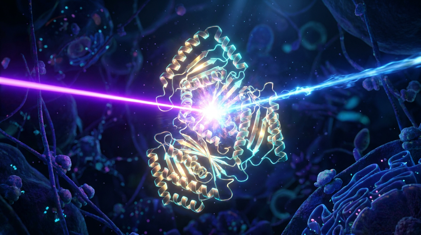
- Researchers successfully demonstrated the technology by imaging hemoglobin, a notoriously difficult small protein.
- The breakthrough could eventually allow scientists to observe more than 50% of proteins in their natural cellular environment.
- The technology is currently in the prototype phase and requires extreme precision to align.
Inside every human cell, tens of thousands of proteins act as the molecular machinery of life—ferrying cargo, repairing DNA, and triggering diseases when they malfunction. For decades, scientists have sought to map these structures to understand human biology and design targeted drugs. Yet, a fundamental physical limitation has kept the vast majority of these proteins hidden in the dark. Now, a landmark engineering breakthrough is poised to turn the lights on.[3][4][7]
In a series of papers published this week, researchers have unveiled a "laser phase plate" that dramatically enhances the vision of cryo-electron microscopes. By intersecting an electron beam with a laser 100 million times brighter than the sun, the technology boosts image contrast enough to reveal proteins previously considered too small to see.[1][2][4]
To understand the magnitude of this advance, one must look at the current gold standard: cryo-electron microscopy (cryo-EM). This technique, which won the Nobel Prize in Chemistry in 2017, involves flash-freezing biological samples and bombarding them with electrons to deduce their atomic structure. It revolutionized structural biology, but it has a severe blind spot.[3][7]
Conventional cryo-EM struggles to image molecules smaller than 70 kilodaltons. Because the electron beam must be kept weak to avoid destroying the delicate biological sample, the resulting images have an incredibly low signal-to-noise ratio. The problem? Approximately 90% of the human proteome consists of proteins smaller than that 70-kilodalton threshold.[2][3][6]

"If you look at all the proteins in a human, they all have various sizes, and all of these proteins are potential disease mechanisms and drug targets," notes Holger Müller, a physicist at UC Berkeley and Lawrence Berkeley National Laboratory. For years, the inability to clearly image these smaller targets has represented an enormous gap in scientific knowledge.[5]
The solution draws on a century-old concept. In the 1930s, Dutch physicist Frits Zernike discovered that passing light through a biological sample shifts its phase slightly—a change that can be converted into visible contrast. His phase-contrast light microscope won a Nobel Prize in 1953. For decades, engineers have tried to adapt Zernike's phase-contrast principle to the electron beams used in cryo-EM, but physical materials placed in the beam's path quickly accumulated static charge, ruining the image.[2][3][4][6]
His phase-contrast light microscope won a Nobel Prize in 1953.
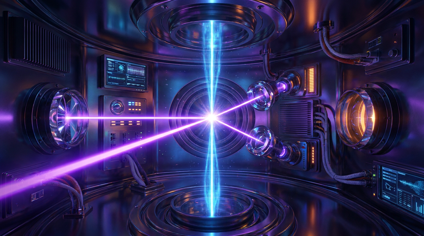
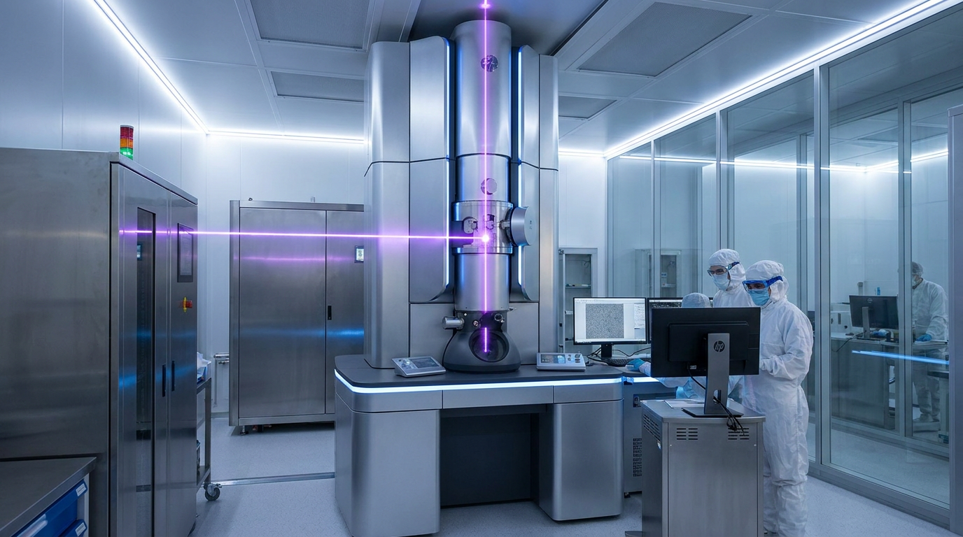
The new approach discards physical materials entirely, using pure light to manipulate the electrons. The engineering required is staggering. The system traps a continuous-wave laser inside a spherical, mirrored cavity, forcing the light to bounce back and forth more than 10,000 times. This concentrates the beam to an intensity of 75 kilowatts within a space of just a few microns.[1][6]
When the microscope's electron beam passes through this intense field of light, the laser acts as an invisible lens. It slows the unscattered electrons just enough to shift their phase by exactly 90 degrees relative to the electrons that scattered off the protein sample. When the two sets of electrons recombine at the detector, they interfere with each other, producing a high-contrast image without requiring a higher, sample-destroying electron dose.[2][4][7]
The evidence for the system's efficacy is robust. In the Science paper, the UC Berkeley team installed the laser phase plate on a customized Thermo Fisher Krios microscope and tested it on hemoglobin, a blood protein that sits at the absolute lower size limit for conventional machines. The laser phase plate significantly improved the resolution of the hemoglobin structure, proving its viability for small targets.[2][3][5]
A parallel effort by the Chan Zuckerberg Biohub pushes the concept even further by developing a dual-laser crossed phase plate. This configuration aims to eliminate the slight optical distortions caused by a single laser beam, paving the way for even higher-fidelity imaging.[4]
Beyond isolated proteins, the technology promises to revolutionize cryo-electron tomography (cryo-ET)—the practice of imaging proteins in their native, crowded cellular environment. Currently, scientists estimate they can image fewer than 1% of proteins in their natural cellular context because cells are too thick and "messy" for conventional electron beams to penetrate cleanly.[3][4]

"It's like a forest of trees, and you're trying to find one leaf on one tree in there," explains Bridget Carragher, founding technical director of imaging at Biohub. By drastically increasing the signal-to-noise ratio, the laser phase plate could eventually make more than 50% of cellular proteins visible in their native state.[4][6]
Despite the excitement, the technology remains in its infancy. Aligning a 75-kilowatt laser cavity inside an electron microscope requires extreme precision, and the systems are currently bespoke prototypes. Furthermore, while the team has successfully imaged proteins down to 50 kilodaltons, they have yet to reach their ultimate theoretical goal of 17 kilodaltons.[2][6][7]

Nevertheless, the successful demonstration of the laser phase plate marks a paradigm shift in structural biology. As the technology is refined and commercialized, it is expected to unlock the structures of thousands of previously invisible proteins, accelerating the discovery of new therapeutics and fundamentally deepening our understanding of the molecular machinery that drives human life.[1][3][7]
How we got here
1930s
Dutch physicist Frits Zernike discovers that shifting the phase of light can create visible contrast in biological samples.
1953
Zernike is awarded the Nobel Prize in Physics for inventing the phase-contrast light microscope.
2017
The Nobel Prize in Chemistry is awarded for the development of cryo-electron microscopy (cryo-EM).
June 2026
Researchers publish successful demonstrations of the laser phase plate, adapting phase-contrast principles to electron beams.
Viewpoints in depth
Structural Biologists' view
A focus on unlocking the 'dark matter' of the human proteome for drug discovery.
For structural biologists, the laser phase plate represents a master key to the 90% of human proteins that are currently too small to image using conventional cryo-EM. Because these sub-70-kilodalton proteins include countless disease mechanisms and drug targets, researchers view the technology as a catalyst for next-generation pharmaceuticals. By finally seeing the exact shape of these molecules, scientists can design drugs that bind to them with unprecedented precision.
Microscopy Engineers' view
A focus on overcoming decades of physical limitations in electron phase shifting.
Engineers have spent decades trying to adapt Frits Zernike's phase-contrast principles to electron microscopes, but physical phase plates inevitably accumulated static charge and ruined the electron beam. The engineering community views the successful deployment of a 75-kilowatt, continuous-wave laser trapped in a mirrored cavity as a monumental triumph of optical physics. The ability to shift an electron beam's phase by exactly 90 degrees using pure light solves a fundamental bottleneck in microscopy hardware.
Cellular Biologists' view
A focus on observing the molecular machinery of life in its native, crowded environment.
While isolating proteins in purified ice is useful, cellular biologists are most excited about the technology's application to cryo-electron tomography (cryo-ET). Cells are incredibly dense and messy, making it nearly impossible to see individual proteins at work. By drastically increasing the signal-to-noise ratio, the laser phase plate promises to let researchers observe proteins interacting in their natural cellular habitats, moving from static snapshots to a dynamic understanding of cellular biology.
What we don't know
- It is unclear how quickly the bespoke laser cavity prototypes can be scaled into commercial microscopes for widespread laboratory use.
- Researchers have not yet reached their theoretical imaging limit of 17 kilodaltons, currently bottoming out around 50 kilodaltons.
- The full impact of the dual-laser crossed phase plate design on optical distortion remains to be extensively tested on complex cellular tissues.
Key terms
- Cryo-electron microscopy (cryo-EM)
- A technique that fires electrons at flash-frozen samples to determine the atomic structure of molecules.
- Proteome
- The entire set of proteins expressed by a genome, cell, tissue, or organism.
- Kilodalton (kDa)
- A unit of mass used to express the molecular weight of proteins.
- Phase shift
- A slight delay in the wave cycle of light or electrons, which can be manipulated to create visual contrast.
- Signal-to-noise ratio
- The measure of true signal (the protein image) compared to background interference (noise).
Frequently asked
Why can't we just use stronger electron beams to see small proteins?
Biological samples are incredibly fragile. A stronger electron beam would destroy the protein before a clear image could be captured.
How does the laser help the electron microscope?
The intense laser acts as an invisible lens, slowing down parts of the electron beam to create a high-contrast image without needing a stronger, destructive dose of electrons.
When will this technology be used for drug discovery?
While the prototypes are currently operational in research labs, it will likely take several years to commercialize the technology for widespread pharmaceutical use.
Sources
Source coverage
7 outlets
3 viewpoints surfaced


[1]NatureStructural Biologists
An innovative technology boosts image quality for protein structures
Read on Nature →[2]ScienceMicroscopy Engineers
Laser phase plate improves structure determination of small proteins by cryo-EM
Read on Science →[3]UC BerkeleyCellular Biologists
Laser phase plate promises to revolutionize cryo-electron microscopy
Read on UC Berkeley →[4]Chan Zuckerberg BiohubCellular Biologists
Making the invisible visible: A landmark achievement in microscopy
Read on Chan Zuckerberg Biohub →[5]News-MedicalStructural Biologists
Laser phase plate could fill enormous gap in knowledge of protein structures
Read on News-Medical →[6]BiocompareMicroscopy Engineers
Laser Phase Plate Enhances Cryo-EM for Small Proteins
Read on Biocompare →[7]Factlen Editorial TeamCellular Biologists
Synthesis by Factlen editorial team
Read on Factlen Editorial Team →

More in science
See all 19 stories →Structural Biology
A 75-Kilowatt Laser Just Unlocked the Invisible World of Human Proteins
0 sources
Wind Power
Renewable Energy Groups Sue Pentagon Over 'De Facto Moratorium' on Wind Farms
0 sources

Cryo-EM Tech
Laser Phase Plate Breakthrough Illuminates the Invisible 90% of the Human Proteome
0 sources
Wind Power Policy
Renewable Energy Groups Sue Pentagon Over 'Total Halt' of Wind Farm Reviews
0 sources

Every angle. Every day.
Get science stories with full source coverage and perspective breakdowns delivered to your inbox.