The Laser Phase Plate: A Century-Old Physics Trick Just Unlocked the Human Proteome
By adapting a 1930s optical breakthrough to modern electron microscopes, physicists have drastically improved our ability to see the tiny proteins that drive human health and disease.
By Factlen Editorial Team
- Structural Biologists
- Focused on mapping the proteome and understanding cellular machinery in its native context.
- Applied Physicists
- Focused on the engineering triumph of manipulating electron waves with concentrated light to avoid electrostatic charging.
- Biomedical Engineers
- Focused on the translation of this technology into drug discovery and targeted therapeutics.
What's not represented
- · Commercial microscope manufacturers scaling the tech
- · Computational biologists analyzing the new data volumes
Why this matters
More than 90% of the proteins in human cells are too small to be clearly imaged by existing cryo-electron microscopes. By bringing these invisible molecular machines into sharp focus, this technology will dramatically accelerate the discovery of new drugs and targeted treatments for diseases.
Key points
- Cryo-electron microscopy struggles to image proteins under 70 kilodaltons, leaving 90% of the human proteome blurry.
- Researchers successfully integrated a 75-kilowatt laser into an electron microscope to act as a phase plate.
- The laser shifts the phase of the electron beam without accumulating static charge, a fatal flaw in physical plates.
- The breakthrough improves the signal-to-noise ratio, allowing clear imaging of proteins as small as 50 kilodaltons.
- The technology paves the way for cryo-electron tomography, enabling scientists to observe proteins inside intact cells.
The interior of a human cell is a crowded, bustling metropolis, but for decades, structural biologists have been trying to map its streets while wearing frosted glasses. Understanding exactly how proteins fold, interact, and misfire is the foundation of modern medicine, yet seeing these mechanisms in action has remained one of biology's most stubborn challenges.[5]
The advent of cryo-electron microscopy (cryo-EM) revolutionized the field, winning a Nobel Prize for its ability to flash-freeze biological samples in vitreous ice and photograph them with a beam of electrons. It allowed scientists to determine the atomic-level architecture of massive molecular machines, accelerating drug discovery and structural biology.[3][4]
But cryo-EM has always suffered from a glaring blind spot. The technique relies on electrons scattering off the atoms in a sample to create an image. Small proteins simply do not scatter enough electrons to stand out from the background noise, leaving them blurry or entirely invisible.[1][5]
The numbers highlight the scale of the problem: proteins under 70 kilodaltons are incredibly difficult to image with conventional electron microscopes. Yet, these diminutive structures account for roughly 90% of the human proteome.[3][4]

This means the vast majority of the molecular machinery that drives human health, cellular function, and disease progression has remained out of reach, forcing scientists to guess at their exact structures or rely on less precise methods.[4]
Enter the laser phase plate. In a series of landmark papers published this week in Science and Nature, researchers from UC Berkeley and the Chan Zuckerberg Biohub have unveiled a solution that finally brings these tiny proteins into sharp focus.[1][2][4]
To understand the mechanics of this breakthrough, it helps to look back nearly a century to the work of Dutch physicist Frits Zernike, who faced a remarkably similar problem with optical light.[3]
In 1932, Zernike realized that when light passes through a transparent biological sample, its phase shifts slightly. By interfering this shifted light with unshifted light, he created stark visual contrast out of thin air—a discovery that won him the 1953 Nobel Prize and transformed light microscopy.[3]
For decades, physicists have desperately wanted to apply Zernike's phase-contrast trick to electron microscopes, knowing it would drastically boost the signal-to-noise ratio for biological samples.[5]
The problem is that electrons, unlike photons, are charged particles. When researchers tried inserting physical phase plates—such as ultra-thin films of carbon—into the electron beam, the plates quickly accumulated static electricity.[5]

The problem is that electrons, unlike photons, are charged particles.
This static charge acted like an erratic, unwanted lens, distorting the electron beam and ruining the image before any useful high-resolution data could be collected.[5]
Physicist Holger Müller and biophysicist Robert Glaeser at UC Berkeley proposed a radical alternative 15 years ago: what if the phase plate wasn't made of physical matter at all?[4]
Their idea was to use light to manipulate electrons. By crossing the electron beam with an incredibly intense laser, the electromagnetic field of the light would shift the phase of the passing electrons without introducing any physical material to hold a static charge.[3][4]
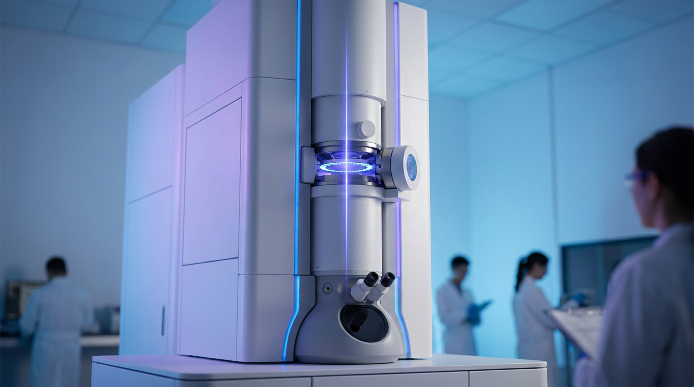
Building the device was an engineering nightmare. The team had to trap a continuous-wave laser inside a spherical, mirrored cavity, bouncing the light back and forth to concentrate 75 kilowatts of power into a beam just a few microns wide.[3]

The grueling effort paid off. Installed in a custom-modified Thermo Fisher Krios microscope, the laser phase plate successfully pulled small, faint proteins out of the background noise and into sharp, high-resolution focus.[3][4]
The team demonstrated the technology on hemoglobin, the oxygen-carrying protein in blood, which sits right at the lower limit of what conventional machines can resolve. The resulting images showed a dramatic improvement in clarity.[2][3]
With the laser phase plate, the signal-to-noise ratio essentially doubled, allowing the researchers to clearly resolve structures as small as 50 kilodaltons, with a theoretical path mapped out to eventually reach 17 kilodaltons.[2][3]
But the ultimate prize of this technology isn't just seeing isolated proteins in a purified solution. It is the advancement of cryo-electron tomography (cryo-ET)—imaging proteins while they are still operating inside intact, living cells.[1][4]

"It's like a forest of trees, and you're trying to find one leaf on one tree in there," explained Bridget Carragher, founding technical director of imaging at Biohub. The laser phase plate provides the contrast needed to cut through that cellular clutter.[4]
History consistently shows that biology follows technology. By finally turning on the lights inside the cell and illuminating the remaining 90% of the proteome, scientists are poised to uncover the exact molecular interactions that trigger diseases, accelerating the next generation of highly targeted therapeutics.[4][5]
How we got here
1932
Frits Zernike discovers phase contrast for light microscopy, later winning the 1953 Nobel Prize.
2010
UC Berkeley physicists first propose the theoretical concept of using a laser as a phase plate for electron microscopes.
2017
Cryo-electron microscopy (cryo-EM) wins the Nobel Prize in Chemistry for high-resolution structure determination of biomolecules.
June 2026
Researchers publish successful demonstrations of the laser phase plate in Science and Nature, breaking the resolution barrier for small proteins.
Viewpoints in depth
Structural Biologists' view
The laser phase plate is the key to observing proteins in their native cellular environments.
For decades, structural biologists have been forced to extract proteins from cells to study them in isolation, stripping away the vital context of how they interact with their surroundings. They view the laser phase plate as the ultimate unlock for cryo-electron tomography (cryo-ET). By providing enough contrast to see small proteins amidst the dense clutter of the cell, this technology will finally allow researchers to observe molecular machinery operating in its native environment.
Applied Physicists' view
This breakthrough is the culmination of a 15-year battle against electrostatic charging in electron optics.
Physicists see this development as a triumph of fundamental engineering over a seemingly insurmountable physical limitation. Because electrons are charged particles, physical phase plates inevitably accumulate static electricity, distorting the beam. By replacing physical matter with a 75-kilowatt photon field trapped in a mirrored cavity, physicists have elegantly solved a fundamental limitation of electron optics that has frustrated the field for half a century.
Pharmaceutical Developers' view
Seeing smaller proteins accelerates the design of targeted drugs for currently untreatable diseases.
The modern drug discovery industry relies heavily on seeing the exact three-dimensional shape of target proteins to design molecules that bind to them perfectly. Because 90% of the human proteome was previously too small to image clearly, pharmaceutical developers view this technology as a massive commercial and medical catalyst. Bringing these 'invisible' proteins into focus will open up entirely new avenues for treating diseases that are currently considered 'undruggable.'
What we don't know
- How quickly the laser phase plate can be commercialized and installed in laboratories worldwide.
- Whether the technology can consistently reach its theoretical resolution limit of 17 kilodaltons.
- How the massive increase in visual data from cryo-ET will be computationally managed and analyzed.
Key terms
- Proteome
- The entire set of proteins that is, or can be, expressed by a genome, cell, tissue, or organism at a certain time.
- Phase contrast
- A technique that translates invisible shifts in the phase of light or electrons into stark visual contrast, making transparent structures visible.
- Cryo-electron tomography (cryo-ET)
- An advanced imaging technique that pieces together multiple 2D images taken from different angles to create a 3D model of a cell's interior.
- Vitreous ice
- Water that has been frozen so rapidly that it doesn't form crystals, preserving biological samples in their natural state without damaging them.
Frequently asked
What is cryo-electron microscopy?
A Nobel-winning technique that flash-freezes biological samples in vitreous ice and uses a beam of electrons to take highly detailed pictures of their molecular structures.
Why couldn't we see small proteins before?
Small proteins do not scatter enough electrons to stand out from the background noise, making them appear blurry or entirely invisible in standard electron microscopes.
What is a kilodalton?
A unit of mass used in biochemistry to measure the size of proteins and molecules. One kilodalton is roughly the mass of 1,000 hydrogen atoms.
How does a laser act as a plate?
Instead of using a physical material that collects static electricity, the system uses the intense electromagnetic field of a concentrated laser to shift the phase of the passing electrons.
Sources
Source coverage
5 outlets
3 viewpoints surfaced


[1]NatureStructural Biologists
An innovative technology boosts image quality for protein structures
Read on Nature →[2]ScienceStructural Biologists
Laser phase plate improves structure determination of small proteins by cryo-EM
Read on Science →[3]UC BerkeleyApplied Physicists
Physicists introduce phase contrast to electron microscopy, delivering sharper images of our body's tiniest proteins
Read on UC Berkeley →[4]Chan Zuckerberg BiohubBiomedical Engineers
Resolving the cell in atomic detail
Read on Chan Zuckerberg Biohub →[5]Factlen Editorial TeamBiomedical Engineers
Synthesis by Factlen editorial team
Read on Factlen Editorial Team →

Every angle. Every day.
Get science stories with full source coverage and perspective breakdowns delivered to your inbox.