A 'Laser Phase Plate' Breakthrough Just Made 90% of Human Proteins Visible to Science
By integrating a laser 100 million times brighter than the sun into an electron microscope, researchers have solved a 15-year physics challenge, allowing scientists to finally see the tiny proteins that drive human health and disease.
By Factlen Editorial Team
- Structural Biologists
- Researchers focused on mapping the fundamental architecture of cellular machinery.
- Pharmaceutical Developers
- Scientists who rely on protein structures to design targeted drugs and therapies.
- Medical Researchers
- Experts studying how proteins interact and malfunction in their native cellular environments to cause disease.
What's not represented
- · Microscope Manufacturers
- · Clinical Trial Investigators
Why this matters
Most modern drugs are designed by looking at the exact 3D shape of a disease-causing protein and building a chemical key to lock it down. By making the smallest, previously invisible proteins clear to see, this technology unlocks thousands of new targets for treating cancer, Alzheimer's, and rare genetic disorders.
Key points
- A new 'laser phase plate' has been successfully integrated into a cryo-electron microscope.
- The device uses a laser 100 million times brighter than the sun to shift the phase of an electron beam.
- This dramatically boosts image contrast, allowing scientists to clearly see small proteins for the first time.
- The breakthrough improves the resolution of hemoglobin, a small blood protein, by up to 44%.
- Researchers estimate the technology will eventually make over 50% of human proteins visible in their native cellular environment.
- The advancement provides a massive boost to structure-based drug discovery for complex diseases.
For the past decade, structural biology has been dominated by a Nobel Prize-winning technology called cryo-electron microscopy (cryo-EM). By flash-freezing biological samples and bombarding them with electrons, scientists have been able to map the intricate 3D shapes of the molecular machines that keep us alive. But cryo-EM has always harbored a massive blind spot: it struggles to see small things. Because biological specimens are 'weak-phase objects' that generate very little intrinsic image contrast, the electron beam passes through them almost unimpeded. As a result, researchers estimate that more than 90% of the proteins inside human cells are simply too small to generate enough contrast to be imaged clearly.[3][4][6]
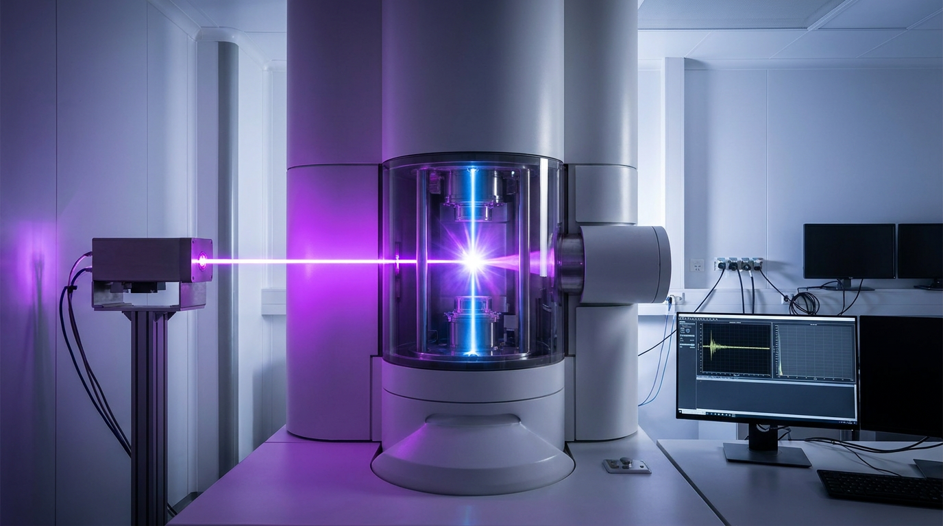
That blind spot is now closing. In a landmark achievement published across Science, Nature Communications, and bioRxiv, researchers from UC Berkeley and Biohub have successfully built and integrated a 'laser phase plate' into a state-of-the-art cryo-electron microscope. First proposed more than 15 years ago by physicists Holger Müller and Robert Glaeser, the device was long dismissed by the field as theoretically elegant but practically impossible to build. Today, it is fully operational, dramatically boosting the contrast of small proteins and bringing the invisible machinery of the cell into sharp focus.[1][2][3][5]

The physics behind the breakthrough relies on manipulating the electron beam itself. Inside the microscope, a powerful laser—amplified by highly polished mirrors to be 100 million times brighter than the surface of the sun—is focused directly onto the electron beam. As the electrons pass through this intense field of light, their phase is shifted. This subtle modulation converts invisible phase shifts into detectable intensity variations when the electrons hit the camera sensor, effectively turning up the contrast dial on the microscopic world.[2][3][4]
To prove the technology works, the Berkeley and Biohub teams tested the laser phase plate on hemoglobin, the oxygen-carrying protein in human blood. At roughly 64 kilodaltons, hemoglobin sits at the absolute lower limit of what conventional cryo-EM can resolve; it typically appears as a blurry, indistinct blob. With the laser switched on, the resolution of the hemoglobin structure improved by up to 44%. Features that were previously washed out sharpened into clearly defined atomic architecture, proving that the laser does not trade resolution for contrast.[2][3][6]

To prove the technology works, the Berkeley and Biohub teams tested the laser phase plate on hemoglobin, the oxygen-carrying protein in human blood.
The teams also achieved a staggering 1.8-angstrom resolution on apoferritin, an iron-storage protein used as a standard benchmark in the field. This approaches the theoretical resolution limit of the technology, signaling to structural biologists that the laser phase plate is not just a proof-of-concept, but a robust tool ready for rigorous scientific application. The device has been successfully integrated into a custom Thermo Scientific Krios microscope, the dominant, multi-million-dollar workhorse of modern structural biology facilities.[1][5][6]
The implications for medicine and drug discovery are profound. The pharmaceutical industry relies heavily on structure-based drug design: if researchers can see the exact geometric shape of a protein that causes a disease, they can design a custom molecule—a ligand or inhibitor—to bind to it and shut it down. Because conventional cryo-EM could only resolve roughly 10% of the human proteome in purified form, thousands of potential drug targets remained locked in a blurry haze. By bringing these elusive proteins into clear view, the laser phase plate provides a direct path toward designing highly specific therapies with fewer off-target side effects.[4][6]

Beyond isolated proteins, the researchers are already looking toward the next frontier: cryo-electron tomography (cryo-ET). While standard cryo-EM requires proteins to be purified and isolated in a solution, cryo-ET captures 3D reconstructions of proteins in their natural, crowded cellular environment. Currently, fewer than 1% of proteins can be seen clearly in their native state. The laser phase plate is expected to push that number past 50%, allowing scientists to watch how molecular machines assemble, interact, and malfunction in real-time disease states.[3][4][6]
While the technology is currently limited to a few specialized laboratories in California, the successful demonstration marks a paradigm shift in biological imaging. By merging quantum optics with structural biology, scientists have finally overcome the contrast barrier that has held the field back for decades. As the laser phase plate moves toward commercialization and broader adoption, it promises to map the fundamental molecular drivers of human life with unprecedented precision.[1][2][6]
How we got here
2010s
Physicists Holger Müller and Robert Glaeser first propose the theoretical concept of a laser phase plate for electron microscopy.
2017
The Nobel Prize in Chemistry is awarded for the development of cryo-electron microscopy, cementing it as the premier tool for structural biology.
Early 2020s
Researchers achieve a working prototype of the laser phase plate in an older-generation electron microscope.
June 11, 2026
UC Berkeley and Biohub publish successful results demonstrating the laser phase plate integrated into a state-of-the-art commercial microscope, achieving record contrast for small proteins.
Viewpoints in depth
Structural Biologists
Researchers focused on mapping the fundamental architecture of life.
For structural biologists, the laser phase plate solves the single most frustrating limitation of their field: the inability to see small, weak-phase objects. While cryo-EM revolutionized the study of massive molecular complexes, the vast majority of cellular machinery remained just out of reach. Biologists view this breakthrough not just as an incremental upgrade, but as a telescope that has finally been focused, allowing them to map the 'dark matter' of the human proteome and observe proteins interacting in their native cellular environments.
Pharmaceutical Developers
Scientists designing the next generation of targeted medicines.
The drug discovery industry relies heavily on structure-based design—literally building chemical keys to fit the specific locks of disease-causing proteins. Because conventional microscopes could not resolve small proteins, many potential drug targets were considered 'undruggable.' Pharmaceutical researchers see the laser phase plate as a massive commercial and medical accelerator. By providing exact geometric maps of previously hidden proteins, the technology will allow for the rapid design of highly specific ligands and inhibitors, reducing off-target side effects and speeding up the development pipeline for complex diseases.
What we don't know
- How quickly the technology can be scaled and commercialized for widespread use in standard research facilities.
- Whether the intense laser field might introduce subtle, unforeseen artifacts when imaging highly delicate cellular structures in tomography.
- The exact timeline for when this enhanced imaging will directly result in new, approved pharmaceutical therapies.
Key terms
- Cryo-electron microscopy (cryo-EM)
- A technique that fires a beam of electrons at flash-frozen biological samples to create high-resolution 3D images of molecules.
- Laser phase plate
- A device that uses an intense, focused laser beam to shift the phase of passing electrons, converting invisible phase differences into high-contrast images.
- Proteome
- The entire set of proteins that is, or can be, expressed by a genome, cell, tissue, or organism at a certain time.
- Cryo-electron tomography (cryo-ET)
- An advanced imaging technique that captures multiple 3D views of proteins in their natural, crowded environment inside an intact cell.
- Angstrom (Å)
- A unit of length equal to one ten-billionth of a meter, commonly used to measure atomic-scale structures.
Frequently asked
Why couldn't we see these proteins before?
Most proteins are very small and generate almost no contrast when an electron beam passes through them, making them appear invisible or highly blurry in standard microscopes.
How does the laser help?
The intense laser light shifts the phase of the electrons as they pass through it. When these electrons hit the camera, that phase shift translates into a sharp, high-contrast image.
Will this cure diseases?
Not directly, but it provides the exact 3D blueprints of the proteins that cause diseases. Drug developers use these blueprints to design highly precise medicines to block or fix those proteins.
Is this technology widely available?
Not yet. It has currently been successfully built and tested in specialized, custom-modified microscopes at UC Berkeley and Biohub, but researchers are working with manufacturers to commercialize it.
Sources
Source coverage
6 outlets
3 viewpoints surfaced


[1]NatureStructural Biologists
An innovative technology boosts image quality for protein structures
Read on Nature →[2]ScienceStructural Biologists
Enhanced contrast for small proteins with the single-laser phase plate
Read on Science →[3]UC BerkeleyMedical Researchers
Microscope Breakthrough Will Open Unprecedented View into Our Cells
Read on UC Berkeley →[4]BiohubMedical Researchers
Making the invisible visible: A landmark achievement in microscopy
Read on Biohub →Theoretical framework for the crossed laser phase plate
Read on Nature Communications →[6]Factlen Editorial TeamPharmaceutical Developers
Synthesis by Factlen editorial team
Read on Factlen Editorial Team →

Every angle. Every day.
Get science stories with full source coverage and perspective breakdowns delivered to your inbox.