Laser Phase Plate Breakthrough Allows Scientists to Image the Cell's Smallest Proteins
By integrating a high-powered laser into cryo-electron microscopes, researchers have solved a decades-old contrast problem, making it possible to visualize tiny, previously invisible proteins that drive human health and disease.
By Factlen Editorial Team
- Structural Biologists
- Focus on the ability to finally visualize the vast majority of the proteome in its native cellular environment rather than in isolated purification.
- Microscopy Engineers
- Emphasize the physical triumph of building a stable 75-kilowatt laser cavity inside an electron microscope to overcome the decades-old electrostatic charging problem.
- Pharmaceutical Researchers
- Value the technology as a catalyst for structure-based drug design, allowing chemists to target disease-causing proteins that were previously invisible.
What's not represented
- · University core facility directors managing equipment budgets
- · Computational biologists developing the software to parse the new data
Why this matters
More than 90% of the proteins that govern human biology and disease are currently too small to be seen clearly by our best microscopes. This breakthrough allows scientists to finally visualize these tiny molecular machines in their natural cellular habitats, dramatically accelerating the discovery of new drugs and targeted therapies.
Key points
- Conventional cryo-electron microscopes struggle to image proteins smaller than 70 kilodaltons, leaving 90% of human proteins out of focus.
- Researchers successfully integrated a 75-kilowatt laser into a microscope to act as a 'phase plate,' boosting image contrast without blurring.
- The laser shifts the phase of the electron beam without relying on physical materials, solving a decades-old electrostatic charging problem.
- The team proved the concept by capturing high-contrast images of hemoglobin, a notoriously difficult 50-kilodalton protein.
- The breakthrough will vastly improve cryo-electron tomography, allowing scientists to map proteins inside intact, living cells.
- Visualizing these previously hidden proteins is expected to dramatically accelerate structure-based drug discovery.
The inner workings of the human cell are driven by a vast, invisible machinery of proteins. For the past decade, a Nobel Prize-winning technology called cryo-electron microscopy (cryo-EM) has allowed scientists to freeze these molecular machines in time and map their atomic structures. But cryo-EM has a glaring blind spot: it struggles to see anything small. Because electrons pass right through tiny proteins without scattering enough to create a visible shadow, the resulting images are washed out and unreadable. Consequently, the vast majority of the human proteome has remained out of focus.[1][5]
That optical barrier has now been broken. In a pair of landmark papers published this week in Science and Nature Communications, researchers from UC Berkeley and the Chan Zuckerberg Biohub announced the successful integration of a "laser phase plate" into a state-of-the-art cryo-electron microscope. By firing a high-intensity laser across the microscope's electron beam, the team successfully manipulated the phase of the electrons, generating unprecedented contrast for biological samples that were previously too faint to image.[1][2][3]
The scale of the problem this solves is immense. Conventional cryo-EM is highly effective for massive molecular complexes, but it hits a practical wall when imaging proteins smaller than roughly 70 kilodaltons. Unfortunately, more than 90 percent of the proteins found inside human cells fall below that threshold. To study them, biologists have historically been forced to pull them out of the cell and crystallize them in isolation, stripping away the crucial context of their natural, crowded environment.[2][5]

To understand the breakthrough, one must understand the physics of contrast. When an electron beam passes through a flash-frozen biological sample, most electrons travel straight through, while a few bounce off the atoms in the protein. To make the protein visible, scientists need those unscattered and scattered electrons to interfere with each other, creating dark and light spots. In standard cryo-EM, operators achieve this by deliberately throwing the microscope slightly out of focus. This "defocusing" creates artificial contrast, but it comes at a steep cost: it blurs the highest-resolution details of the image.[2][6]
The ideal solution has been known in optical physics for nearly a century. In the 1930s, Dutch physicist Frits Zernike won a Nobel Prize for inventing the phase-contrast light microscope, which uses a physical glass plate to shift the phase of unscattered light, creating sharp contrast without blurring the focus. For decades, microscopy engineers have tried to replicate Zernike's physical phase plate for electron microscopes. However, inserting a physical object into an electron beam causes the material to rapidly accumulate a static electrical charge, which wildly distorts the image and renders the technique useless.[2][4]
The UC Berkeley team, led by physicist Holger Müller, realized they needed a phase plate made of something that couldn't hold an electrical charge: light itself. First proposed theoretically more than 15 years ago, the concept involves using the electromagnetic field of a tightly focused laser to slow down the electrons just enough to shift their phase by exactly 90 degrees. While theoretically elegant, building a laser intense enough to affect electrons traveling at near light-speed inside the cramped, vacuum-sealed column of an electron microscope was considered an extreme engineering long-shot.[2][5]
The UC Berkeley team, led by physicist Holger Müller, realized they needed a phase plate made of something that couldn't hold an electrical charge: light itself.
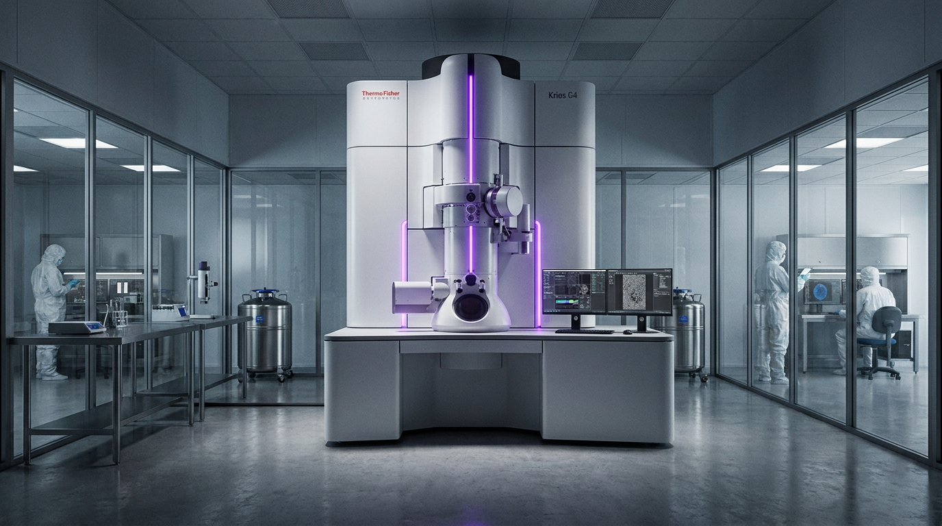
To achieve it, the researchers built a customized mirrored cavity inside a Thermo Fisher Krios microscope. They pumped in a continuous-wave laser and trapped the light, bouncing it back and forth until it reached an astonishing 75 kilowatts of circulating power—concentrated into a beam just a few micrometers wide. When the unscattered electrons pass through this intense wall of light, they experience the necessary phase shift without ever touching a physical surface, completely eliminating the electrostatic charging problem.[2][6]

The results have stunned the structural biology community. To prove the system's efficacy, the researchers targeted hemoglobin, the oxygen-carrying protein in red blood cells. At roughly 50 kilodaltons, hemoglobin sits well below the reliable threshold for conventional cryo-EM and is notoriously difficult to image. With the laser phase plate engaged, the contrast jumped dramatically, allowing the team to reconstruct the protein's structure with remarkable clarity. The smaller the sample, the researchers noted, the more pronounced the improvement provided by the laser.[2][5]
Beyond single-particle imaging, the laser phase plate is poised to revolutionize an emerging field called cryo-electron tomography (cryo-ET). Unlike standard cryo-EM, which images purified proteins in a thin layer of ice, cryo-ET takes multiple angular snapshots of an intact cell to build a 3D map of its interior. Because cells are thick and densely packed, the signal-to-noise ratio in cryo-ET is notoriously poor. The massive contrast boost from the laser phase plate acts like a fog-light, cutting through the cellular noise to reveal individual proteins operating in their native environments.[3][6]
The implications for pharmaceutical research are profound. Currently, drug developers can only use cryo-EM to visualize about 10 percent of the human proteome in purified form, and less than 1 percent in its native cellular context. With the laser phase plate, researchers estimate they will eventually be able to visualize more than 50 percent of the proteins that carry out cellular functions. Seeing exactly how a disease-causing protein folds and interacts with its neighbors inside a living cell allows chemists to design highly specific drugs that lock into the protein's structure like a key in a keyhole.[3][5]

Despite the triumph, the technology remains in its infancy. The current prototype is a bespoke, highly complex modification of an already multi-million-dollar instrument. Maintaining the precise alignment of a 75-kilowatt laser cavity inside a microscope that is highly sensitive to the slightest vibration or temperature change requires constant expert tuning. It will likely be several years before laser phase plates become a commercial add-on available to standard university core facilities.[2][3]
Furthermore, while the hardware has taken a massive leap forward, the software must now catch up. As the laser phase plate illuminates the densely packed "messiness" of intact cells, biologists are suddenly faced with an overwhelming amount of visual data. Identifying specific proteins within a crowded tomogram remains a computational bottleneck, requiring new advances in artificial intelligence and machine learning to parse the newly visible cellular landscapes.[3][6]
Nevertheless, the successful demonstration of the laser phase plate marks a definitive turning point in structural biology. By solving the fundamental physics problem of electron phase contrast, researchers have opened a new window into the microscopic world. The ability to watch the cell's smallest machines at work, in sharp focus and in their natural habitat, promises to rewrite our understanding of biology from the atoms up.[1][3][5]
How we got here
1953
Frits Zernike wins the Nobel Prize in Physics for inventing the phase-contrast optical microscope.
2010
Physicists first propose the theoretical concept of using a focused continuous-wave laser as a phase plate for electron microscopes.
2017
Cryo-electron microscopy (cryo-EM) developers are awarded the Nobel Prize in Chemistry for high-resolution structure determination of biomolecules.
June 2026
UC Berkeley and Biohub researchers publish successful demonstrations of the laser phase plate in Science and Nature Communications.
Viewpoints in depth
Structural Biologists
Focus on the ability to finally visualize the vast majority of the proteome in its native cellular environment rather than in isolated purification.
For decades, structural biologists have had to compromise. To get a clear picture of a protein, they had to remove it from the cell, purify it, and freeze it in isolation. This process strips away the vital context of how the protein interacts with its neighbors. Biologists view the laser phase plate as the key to unlocking cryo-electron tomography (cryo-ET), which allows them to map the cell's interior intact. By boosting the contrast of these "messy" cellular environments, they hope to transition from studying isolated molecular parts to observing the entire cellular engine running in its native state.
Microscopy Engineers
Emphasize the physical triumph of building a stable 75-kilowatt laser cavity inside an electron microscope to overcome the decades-old electrostatic charging problem.
From an engineering perspective, the laser phase plate is a triumph over extreme physical constraints. Previous attempts to create phase contrast in electron microscopes relied on physical carbon films, which quickly accumulated static electricity from the electron beam, destroying the image. Engineers view the laser solution—using the electromagnetic field of light itself to shift the electrons—as an elegant workaround. The primary achievement was designing a mirrored cavity capable of trapping 75 kilowatts of laser power in a space just micrometers wide, all while maintaining perfect alignment inside the vacuum of a highly sensitive microscope.
Pharmaceutical Researchers
Value the technology as a catalyst for structure-based drug design, allowing chemists to target disease-causing proteins that were previously invisible.
The pharmaceutical industry relies heavily on structure-based drug design—creating chemical compounds that perfectly fit into the physical pockets of disease-causing proteins. However, if a protein is too small to image, chemists are essentially designing drugs in the dark. Pharmaceutical researchers see the laser phase plate as a massive expansion of their target catalog. By lowering the visibility threshold from 70 kilodaltons down to 50 or even 17 kilodaltons, the technology exposes thousands of previously hidden human proteins, opening up entirely new avenues for targeted therapies and accelerating the drug discovery pipeline.
What we don't know
- Whether the technology can reliably be pushed down to the ultimate theoretical goal of imaging 17-kilodalton proteins.
- How quickly the computational software will advance to handle the massive influx of complex visual data generated by high-contrast cellular tomograms.
- When, or if, the highly customized and expensive laser phase plate setups can be commercialized and scaled for widespread use in standard research laboratories.
Key terms
- Cryo-electron microscopy (Cryo-EM)
- A technique that fires a beam of electrons through flash-frozen biological samples to determine the 3D atomic structure of molecules.
- Cryo-electron tomography (Cryo-ET)
- An advanced form of cryo-EM that takes multiple pictures of an intact cell from different angles to build a 3D map of its internal environment.
- Phase contrast
- An optical technique that converts invisible shifts in a wave's phase (caused by passing through a transparent object) into visible changes in brightness.
- Proteome
- The entire set of proteins that is, or can be, expressed by a genome, cell, tissue, or organism at a certain time.
- Kilodalton (kDa)
- A unit of mass used to express molecular weight, particularly for large biological molecules like proteins.
Frequently asked
Why can't we use regular light microscopes to see proteins?
Proteins are much smaller than the wavelength of visible light, making them physically impossible to resolve with optical microscopes. Scientists must use electrons, which have much shorter wavelengths, to see atomic-level details.
What exactly does a phase plate do?
It slows down the electrons that pass straight through a sample without hitting anything, shifting their wave pattern. When these shifted electrons recombine with the electrons that did hit the sample, they create a high-contrast image.
Why use a laser instead of a glass lens?
Putting a physical object like a glass or carbon plate into an electron beam causes it to rapidly build up static electricity, which distorts the image. A laser provides the necessary electromagnetic field to shift the electrons without any physical material to hold a charge.
Will this cure diseases immediately?
No, but it provides a crucial foundational tool. By allowing scientists to see exactly what disease-causing proteins look like, it significantly speeds up the process of designing drugs that can bind to and disable them.
Sources
Source coverage
6 outlets
3 viewpoints surfaced


[1]NatureStructural Biologists
An innovative technology boosts image quality for protein structures
Read on Nature →[2]UC BerkeleyMicroscopy Engineers
Physicists introduce phase contrast to electron microscopy, delivering sharper images of our body's tiniest proteins
Read on UC Berkeley →[3]CZ BiohubStructural Biologists
Resolving the cell in atomic detail with laser phase plates
Read on CZ Biohub →[4]Phys.orgMicroscopy Engineers
Physicists introduce phase contrast to electron microscopy, delivering sharper images of our body's tiniest proteins
Read on Phys.org →[5]News-MedicalPharmaceutical Researchers
Laser phase plate dramatically improves contrast of images produced by cryo-electron microscopes
Read on News-Medical →[6]Lawrence Berkeley National LaboratoryMicroscopy Engineers
A Laser-Powered Leap for Cryo-Electron Microscopy
Read on Lawrence Berkeley National Laboratory →

Every angle. Every day.
Get science stories with full source coverage and perspective breakdowns delivered to your inbox.