A 'Laser Phase Plate' Breakthrough Allows Cryo-EM to Image the Tiniest Human Proteins
Researchers have successfully integrated a high-intensity laser into an electron microscope, solving an 80-year contrast problem and opening the door to imaging the 90% of human proteins previously too small to see.
By Factlen Editorial Team
- Structural Biologists
- View the technology as a long-awaited solution to the contrast limits of cryo-EM, unlocking the vast majority of the human proteome for drug discovery.
- Microscope Engineers
- Focus on the immense technical triumph of stabilizing a high-intensity laser cavity inside an electron beam vacuum after 15 years of theoretical doubt.
- Biomedical Researchers
- Anticipate that the technology will revolutionize cryo-electron tomography, allowing them to observe proteins interacting in their natural cellular environments.
What's not represented
- · Commercial Microscope Manufacturers
- · Computational Biologists
Why this matters
Because proteins are the molecular machines that drive human health and disease, seeing their exact 3D structure is the first step in designing new life-saving drugs. This technology makes the vast majority of the human proteome visible to structural biologists for the first time.
Key points
- Conventional cryo-EM struggles to image small proteins, leaving 90% of the human proteome difficult to resolve.
- Researchers have successfully built a 'laser phase plate' that uses intense light to shift the electron beam's phase.
- The device dramatically increases image contrast without relying on physical materials that degrade or contaminate.
- In paired tests, the laser improved the resolution of the hemoglobin protein by up to 44%.
- The breakthrough paves the way for imaging smaller drug targets and viewing proteins in their natural cellular environments.
For decades, structural biologists have relied on cryo-electron microscopy (cryo-EM) to map the molecular machinery of life. The technique, which won the 2017 Nobel Prize in Chemistry, fires electrons at flash-frozen biological samples to deduce their three-dimensional shapes. Yet, despite its revolutionary impact on drug discovery, conventional cryo-EM harbors a massive blind spot: it struggles to generate enough image contrast to resolve small molecules. Researchers estimate that more than 90% of the proteins inside a human cell are simply too small to be imaged clearly using standard methods.[5][6]
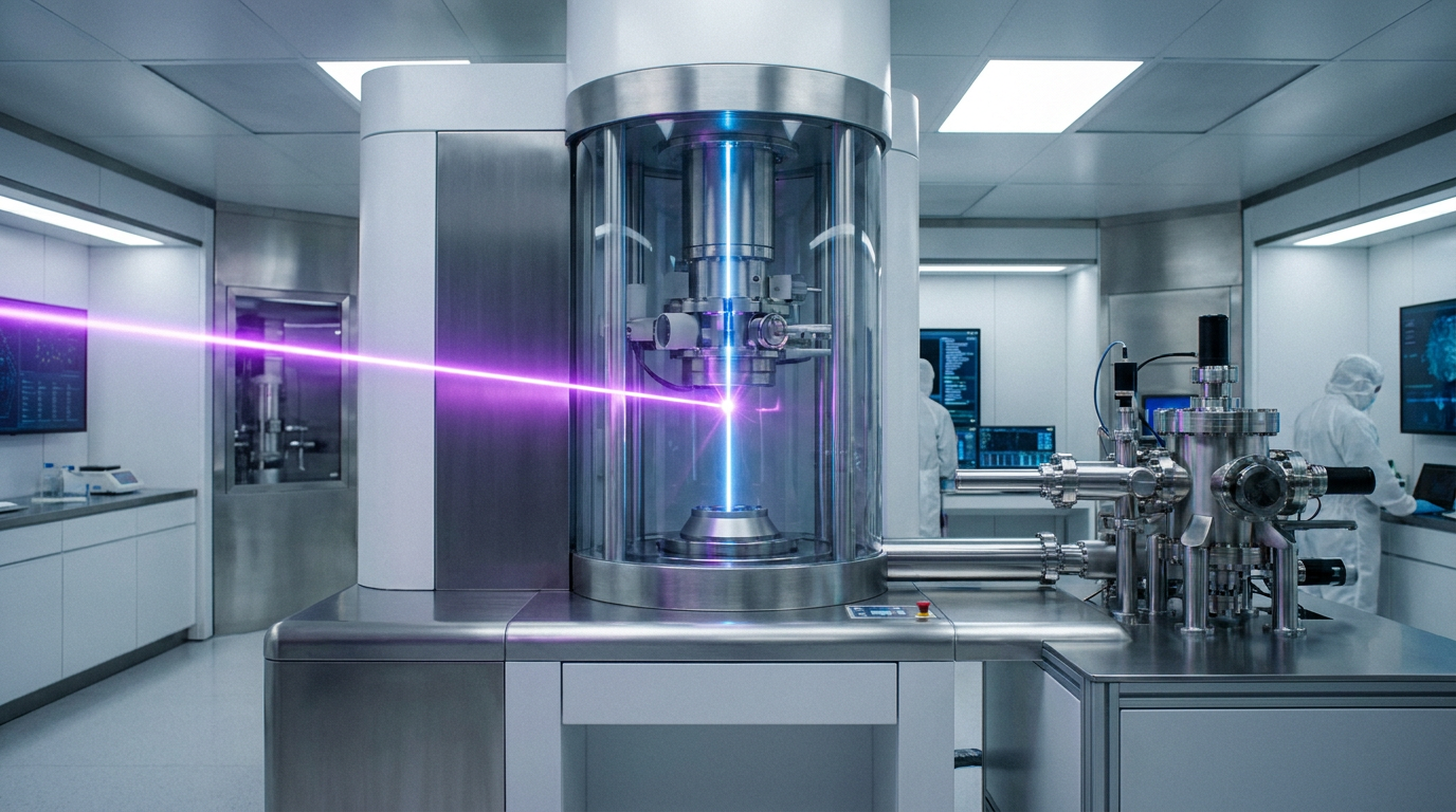
That visibility gap is now closing. In a coordinated suite of publications released this week, researchers from UC Berkeley and the Chan Zuckerberg Biohub announced the successful demonstration of a "laser phase plate" (LPP). Installed inside a state-of-the-art electron microscope, the device uses a highly focused laser to shift the phase of the electron beam, dramatically boosting image contrast without destroying the high-resolution signal.[3][5]
The underlying physics borrows from a nearly century-old optical breakthrough. In the 1930s, the invention of the phase-contrast light microscope allowed biologists to see transparent cellular structures that were previously invisible, earning a Nobel Prize in 1953. Translating that concept to electron microscopes, however, proved notoriously difficult. Physical phase plates made of carbon or other materials tend to charge up, contaminate quickly, and degrade the electron beam, compromising the very atomic-level resolution scientists seek.[4][5]

The solution, first proposed more than 15 years ago by UC Berkeley physicists Holger Müller and Robert Glaeser, was to replace the physical material entirely with light. Their theoretical framework suggested that focusing a continuous-wave laser at the exact point where the electron beam passes could shift the electrons' phase via the ponderomotive effect—with nothing solid in the beam path to burn or contaminate.[3][5]
For years, the concept was dismissed by many in the field as theoretically elegant but practically impossible to build. Generating the necessary phase shift requires a laser beam roughly 100 million times brighter than the surface of the sun, all contained within the ultra-high vacuum of an electron microscope column. Furthermore, the optical cavity must remain perfectly aligned at the microscopic level while avoiding any dust particles that could absorb the intense light and instantly vaporize the mirrors.[3][5]
For years, the concept was dismissed by many in the field as theoretically elegant but practically impossible to build.
The new publications confirm that the engineering hurdles have been cleared. Writing in the journal Science, Müller’s team detailed the performance of a custom Thermo Scientific Krios microscope—nicknamed "Theia"—outfitted with the LPP. The entire laser apparatus fits inside a device under four inches wide, dropping seamlessly into the microscope's column so that, from the outside, the instrument looks like any other standard cryo-EM setup.[1][4]

The evidence of the LPP's efficacy is striking. The Science paper presents paired laser-on and laser-off experiments using several biological benchmarks. For aldolase, a standard benchmark enzyme, the LPP provided a clear boost in contrast. More critically, the team successfully imaged hemoglobin, a 64-kilodalton blood protein that sits at the absolute lower frontier of what conventional cryo-EM can resolve.[1][3]
With the laser engaged, the resolution of the hemoglobin structure improved by up to 44%. Features that appeared as blurry blobs in standard imaging sharpened into clearly defined atomic structures. The team also imaged apoferritin at a resolution of 1.8 angstroms, approaching the theoretical limit of the technology and proving that the laser does not trade away high-resolution detail to achieve its contrast boost.[3]

A parallel effort at CZ Biohub is already pushing the technology further. Described in a preprint and supported by theoretical work published in Nature Communications, the Biohub team has developed a dual-laser system. By crossing two perpendicular laser beams operating at half power, this next-generation design reduces optical aberrations and makes the delicate mirror components significantly less likely to burn out during sustained use.[3][7]
Despite the triumphant data, the researchers emphasize transparent uncertainty regarding the timeline for widespread adoption. The current LPP systems are bespoke prototypes operating in highly specialized physics labs. For the technology to reach the thousands of structural biology core facilities worldwide, it must be commercialized and scaled by major microscope manufacturers, a process that will require substantial industrial engineering.[3]
Furthermore, while the LPP dramatically improves the imaging of purified proteins, its ultimate promise lies in cryo-electron tomography (cryo-ET)—the practice of imaging proteins in their natural, crowded cellular environment. Conventional cryo-ET suffers from notoriously low signal-to-noise ratios. The LPP's contrast boost is expected to be a game-changer for tomography, but extensive in-situ cellular imaging with the new plate remains an active area of development.[4][5]
If successfully commercialized, the laser phase plate will fundamentally alter the landscape of drug discovery. Most modern pharmaceuticals are designed by finding a small molecule that fits perfectly into a specific pocket on a disease-causing protein. If 90% of those proteins cannot be seen clearly, they cannot be effectively targeted. By making the invisible visible, the LPP provides the foundational blueprints needed to design the next generation of targeted therapeutics.[3][5]
How we got here
1930s
The phase-contrast light microscope is invented, eventually winning the 1953 Nobel Prize in Physics.
2010
Holger Müller and Robert Glaeser publish the theoretical framework for using a laser to shift the phase of an electron beam.
2017
The Nobel Prize in Chemistry is awarded to the pioneers of cryo-electron microscopy (cryo-EM).
June 2026
Researchers at UC Berkeley and CZ Biohub publish the first successful demonstrations of a working laser phase plate in a modern cryo-EM system.
Viewpoints in depth
Structural Biologists
View the technology as a long-awaited solution to the contrast limits of cryo-EM.
For structural biologists, the laser phase plate represents the clearing of a decades-old bottleneck. While the 'resolution revolution' of the 2010s allowed cryo-EM to map massive macromolecular complexes, the everyday proteins that drive cellular metabolism and disease remained frustratingly out of reach. By artificially boosting contrast, the LPP allows researchers to apply existing single-particle reconstruction software to molecules as small as 64 kilodaltons. This immediately expands the pool of viable drug targets, allowing pharmaceutical researchers to design inhibitors for proteins that were previously invisible.
Microscope Engineers
Focus on the immense technical triumph of stabilizing a high-intensity laser cavity.
From an engineering standpoint, the LPP was long considered a pipe dream. Early physical phase plates made of carbon film suffered from rapid degradation, charging effects, and signal loss. The theoretical alternative—using light to shift the electron phase—required focusing a continuous-wave laser to an intensity 100 million times brighter than the sun, all within a four-inch device operating in a high vacuum. Engineers celebrate the LPP not just for its biological output, but as a masterclass in optical precision, overcoming thermal instability and mirror degradation to create a stable, usable instrument.
What we don't know
- How quickly commercial microscope manufacturers can scale the delicate laser cavity design for mass production and routine laboratory maintenance.
- Whether the dual-laser system will completely eliminate the risk of mirror burnout during the high-throughput, 24/7 imaging cycles typical of pharmaceutical research.
- The exact lower size limit of proteins that can be resolved using the LPP, as researchers continue to push the boundaries below 64 kilodaltons.
Key terms
- Cryo-electron microscopy (cryo-EM)
- An imaging technique that fires a beam of electrons at flash-frozen samples to map the 3D structure of biological molecules.
- Phase contrast
- An optical technique that translates invisible phase shifts in waves (like light or electrons) into visible changes in brightness and contrast.
- Proteome
- The entire set of proteins expressed by a genome, cell, tissue, or organism at a certain time.
- Cryo-electron tomography (cryo-ET)
- An advanced imaging technique that captures multiple 2D images of a sample from different angles to reconstruct a 3D model of a cell's interior.
- Angstrom (Å)
- A unit of length equal to one ten-billionth of a meter, commonly used to express the size of atoms and molecules.
Frequently asked
What is cryo-electron microscopy (cryo-EM)?
Cryo-EM is a Nobel Prize-winning technique that fires electrons at flash-frozen biological samples to determine the 3D atomic structure of molecules.
Why do we need a laser phase plate?
Conventional cryo-EM lacks the contrast needed to clearly image very small molecules. The laser phase plate boosts this contrast, making the remaining 90% of human proteins visible.
How does the laser improve the image?
The intense laser beam shifts the phase of the electrons as they pass through the sample. When these shifted electrons interact with unscattered electrons, they create a high-contrast interference pattern without damaging the high-resolution data.
Is this technology available in all labs now?
Not yet. The current systems are custom-built prototypes. It will take time for major microscope manufacturers to commercialize the technology for widespread laboratory use.
Sources
Source coverage
7 outlets
3 viewpoints surfaced


[1]ScienceStructural Biologists
Laser phase plate improves structure determination of small proteins by cryo-EM
Read on Science →[2]NatureStructural Biologists
An innovative technology boosts image quality for protein structures
Read on Nature →[3]LabcriticsMicroscope Engineers
Laser Phase Plate Becomes Reality: Biohub and Berkeley Solve Cryo-EM's 80-Year Contrast Problem with a Laser
Read on Labcritics →[4]Phys.orgBiomedical Researchers
Physicists introduce phase contrast to electron microscopy, delivering sharper images of our body's tiniest proteins
Read on Phys.org →[5]Chan Zuckerberg BiohubBiomedical Researchers
Microscope Breakthrough Will Open Unprecedented View into Our Cells
Read on Chan Zuckerberg Biohub →[6]News-MedicalStructural Biologists
Laser phase plate dramatically expands cryo-EM imaging capabilities
Read on News-Medical →Crossed laser phase plates for transmission electron microscopy
Read on Nature Communications →

Every angle. Every day.
Get science stories with full source coverage and perspective breakdowns delivered to your inbox.