
New Laser Technique Measures Quantum Forces in Proteins, Revolutionizing Precision Drug Design
Researchers have developed a label-free laser technique that directly measures the quantum forces shaping proteins in real time. By capturing these elusive interactions, the method promises to dramatically accelerate precision drug design and antiviral screening.
By Logan Price

- Biophysicists & Researchers
- Focus on the ability to directly measure noncovalent quantum forces in real time.
- Pharmaceutical Developers
- Prioritize the technique's potential to replace brute-force drug screening with rapid, predictive models.
- Clinical Diagnosticians
- Interested in the label-free, minimal-preparation aspect of TRIP for rapid patient testing.
Fast facts
- A new laser technique called TRIP directly measures the quantum forces holding proteins together.
- The method bypasses previous thermal barriers by cooling the substrate to near-freezing temperatures.
- TRIP successfully mapped how the SARS-CoV-2 main protease physically rearranged itself.
- The technique accurately predicted the binding affinity of antiviral drugs in real time.
- TRIP operates without invasive chemical labels and requires minimal sample preparation.
- The breakthrough could replace slow, brute-force drug screening with rapid, automated quantum measurement.
Why this matters
By allowing scientists to directly observe how drugs bind to proteins in real time, this technique could replace years of trial-and-error drug screening with rapid, precise measurements. This means faster development of targeted therapies and antivirals for emerging diseases.
How we got here
1960s–2010s
Biologists theorize the role of quantum forces like pi-pi stacking in protein folding, but lack tools to measure them directly in aqueous environments.
December 2002
Early experiments use powerful lasers to measure protein flexibility, but struggle with heat damage and resolution limits.
July 2023
Texas A&M researchers publish initial proof-of-concept for TRIP in PNAS, demonstrating label-free screening on cooled substrates.
June 2026
The team publishes a breakthrough in Science Advances, successfully using TRIP to directly measure quantum forces in the SARS-CoV-2 main protease.
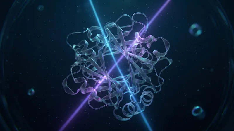
Researchers have solved a decades-old biophysics problem by inventing a laser technique that directly measures the quantum forces holding proteins together. The method, called Thermostable Raman Interaction Profiling (TRIP), allows scientists to watch exactly how pharmaceutical drugs bind to targets like the SARS-CoV-2 virus in real time, without destroying the sample. By capturing these elusive interactions, the technique promises to dramatically accelerate precision drug design and antiviral screening, moving the pharmaceutical industry away from brute-force trial and error toward highly targeted, empirical quantum measurement. The fundamental machinery of life operates on a molecular scale, where proteins twist, fold, and interact to drive everything from immune responses to digestion and cellular repair. For decades, scientists have known that these complex biological interactions are governed by subtle quantum mechanical forces, but observing them directly remained impossible.[1][2][3]
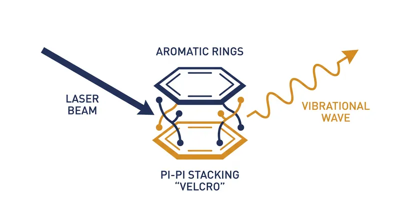
One of the most critical of these quantum forces is aromatic pi-pi stacking. Often conceptualized by structural biologists as biology's 'molecular Velcro,' pi-pi stacking involves the noncovalent attractive forces generated by delocalized electron clouds in flat, ring-shaped molecules. When these aromatic rings approach one another, their electron clouds interlock, creating a stabilizing force that is essential for maintaining the structural integrity of complex biological molecules. These pi-pi stacking forces act as a structural cornerstone across the life sciences. They govern the three-dimensional architecture of folded proteins, maintain the stability of DNA double helices, and dictate the binding affinity of small-molecule inhibitors to therapeutic targets. In the context of pharmaceutical development, the strength and geometry of these stacking interactions often determine whether a drug will successfully bind to a disease-causing protein or simply bounce off.[2][3]
Yet, despite their undeniable importance, measuring these forces directly in living, aqueous systems has historically been impossible. Drug designers and structural biologists were effectively working blindfolded, relying on visual intuition and indirect inference rather than direct empirical measurement. The inability to observe these noncovalent quantum mechanical forces in real time meant that pharmaceutical companies often had to resort to massive, high-throughput screening campaigns—testing thousands of chemical compounds in the hopes of finding one that happened to interact correctly with the target protein. Traditional modalities used to study protein structures have significant limitations. X-ray crystallography, the long-standing gold standard for structural biology, is restricted to static, frozen crystalline states that do not accurately reflect how proteins behave in a fluid, dynamic environment.[1][3]
Similarly, cryo-electron microscopy requires flash-frozen samples, while standard fluorescence or UV spectroscopy relies on invasive chemical labels that risk perturbing the native molecular geometry of the protein. None of these methods can capture the real-time quantum forces at play during a binding event. Attempting to use standard Raman spectroscopy—which fires lasers at a sample to measure vibrational frequencies—presented an entirely different problem. The intense heat generated by the monochromatic light rapidly denatured the fragile proteins before useful, reproducible data could be extracted. For more than half a century, this thermal barrier frustrated biomedical researchers, rendering Raman spectroscopy largely ineffective for studying live, delicate biological molecules in their native states. The heat simply destroyed the very interactions the scientists were attempting to measure.[3][4]
None of these methods can capture the real-time quantum forces at play during a binding event.
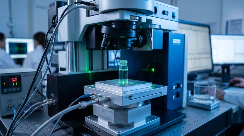
A multidisciplinary team at Texas A&M University finally bypassed this thermal barrier by engineering the TRIP platform. By precisely cooling the substrate to between 0°C and 5°C, they stabilized the proteins, allowing them to probe the samples with high-resolution Raman spectroscopy without causing heat damage. This thermostable environment keeps the proteins intact and fully functional, enabling the laser to penetrate the aqueous solution and extract highly reproducible, label-free data about the molecular vibrations occurring within the sample. When the targeted laser hits the cooled liquid solution, it induces microscopic vibrations across specific chemical bonds. The researchers discovered that a singular spectroscopic marker—the 'Benzene Ring Breathing' vibration mode inherent to the amino acid phenylalanine—functions as a highly sensitive reporter of localized pi-pi stacking.[1][4][5]
As the ring-shaped structures approach one another and stack into parallel or offset configurations, the resulting shift in their vibrational frequency provides a direct, quantifiable readout of the quantum mechanical forces at play. To validate the technique, the team tested TRIP on the main protease (Mpro) of the SARS-CoV-2 virus, a protein that relies heavily on pi-pi stacking for its structural integrity and replication. The empirical data was definitive: the laser not only mapped how the viral protein physically rearranged itself but accurately predicted how effectively various antiviral drugs would bind to it. By tracking the Benzene Ring Breathing mode, the researchers could literally watch the molecular Velcro engage, providing a real-time assessment of drug potency.[1][2][3]
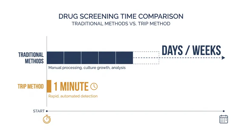
The evidence shows that TRIP can evaluate protein-ligand interactions in as little as one minute, operating at concentrations as low as 1 micromolar. This represents a massive leap in efficiency over traditional methods, which often require days or weeks of sample preparation and analysis. However, the current data relies heavily on the presence of specific aromatic amino acids to generate the necessary Raman signal. It remains an open question how effectively the technique can map forces in proteins lacking these ring structures, or how it will perform in the chaotic, multi-protein environment of a whole cell rather than an isolated laboratory solution. Researchers must also determine if the cooling process itself subtly alters the binding kinetics compared to normal human body temperatures.[4][5][6]
By transitioning structural biology from static snapshots to the direct tracking of quantum mechanical forces, TRIP offers a new automated protocol for drug discovery. If scalable, this label-free optical platform could dramatically accelerate the prescreening of precision therapies, moving the pharmaceutical industry away from brute-force trial and error. As researchers explore extending the technique to analyze DNA and other biological components, TRIP stands poised to become a foundational tool in both clinical diagnostics and next-generation pharmaceutical engineering. The ability to rapidly identify specific protein interactions in a clinical setting could drastically reduce the time patients wait for accurate test results during viral outbreaks, marking a significant leap forward in precision medicine.[1][3][6]
Viewpoints in depth
Biophysicists & Researchers
Focus on the ability to directly measure noncovalent quantum forces in real time.
For decades, structural biologists have relied on static snapshots from X-ray crystallography or cryo-electron microscopy to infer how proteins interact. The introduction of TRIP allows researchers to move beyond visual intuition and directly track the quantum mechanical forces—specifically pi-pi stacking—that dictate molecular behavior. By capturing the 'Benzene Ring Breathing' mode in real time, biophysicists can now observe the dynamic, native-like conditions of proteins without the need for invasive chemical labels.
Pharmaceutical Developers
Prioritize the technique's potential to replace brute-force drug screening with rapid, predictive models.
The traditional drug discovery pipeline is notoriously slow and expensive, often relying on brute-force screening where thousands of compounds are tested to find a viable candidate. Pharmaceutical developers view TRIP as a paradigm-shifting tool that could automate and accelerate this process. Because the technique can evaluate protein-ligand binding affinity in as little as one minute using minimal sample volumes, it offers a highly efficient, cost-effective method for prescreening precision therapies before they enter clinical trials.
Clinical Diagnosticians
Interested in the label-free, minimal-preparation aspect of TRIP for rapid patient testing.
Beyond drug development, the clinical applications of TRIP present a significant opportunity for diagnostic medicine. Because the Raman spectroscopy method requires little to no sample preparation and operates in near-physiological aqueous conditions, diagnosticians see its potential for same-day viral detection. The ability to rapidly identify specific protein interactions in a clinical setting could drastically reduce the time patients wait for accurate test results during outbreaks.
Key terms
- Thermostable Raman Interaction Profiling (TRIP)
- A label-free optical technique that uses a laser on a cooled substrate to measure molecular vibrations and quantum forces in proteins.
- Pi-pi stacking
- A noncovalent attractive force between flat, ring-shaped molecules, often described as biology's 'molecular Velcro.'
- Raman spectroscopy
- A chemical analysis method that shines monochromatic light onto a sample and records the scattered light to identify molecular vibrations.
- Benzene Ring Breathing
- A specific vibrational signature of aromatic amino acids that acts as a sensitive reporter for quantum mechanical interactions.
- Ligand
- A molecule, such as a pharmaceutical drug, that binds to a specific target protein to alter its function.
What we don’t know
- Whether the 'Benzene Ring Breathing' mode can be reliably used to map interactions in proteins that lack high concentrations of aromatic amino acids.
- How the technique will scale from isolated protein-ligand solutions in a controlled lab to the chaotic, multi-protein environment of a living cell.
- The exact timeline for commercializing the TRIP platform into a standard high-throughput screening tool for pharmaceutical companies.
Sources
Source coverage
6 outlets
3 viewpoints surfaced





[1]Texas A&M UniversityBiophysicists & Researchers
Scientists measure hidden quantum forces that could power a new generation of pharmaceutical drugs
Read on Texas A&M University →[2]Quantum ZeitgeistPharmaceutical Developers
TRIP Validated by Predicting Coronavirus Protein Behavior
Read on Quantum Zeitgeist →[3]Quantum Computing ReportPharmaceutical Developers
Capturing Biology's Quantum Velcro via the Benzene Ring Breathing Mode
Read on Quantum Computing Report →[4]Syntec OpticsClinical Diagnosticians
Thermostable Raman Interaction Profiling (TRIP)
Read on Syntec Optics →[5]PNASBiophysicists & Researchers
Thermostable Raman interaction profiling (TRIP) technique that facilitates low-concentration and low-dose screening of binding between protein and ligand
Read on PNAS →[6]Factlen Editorial TeamBiophysicists & Researchers
Synthesis by Factlen editorial team
Read on Factlen Editorial Team →
Comments

Every angle. Every day.
Get science stories with full source coverage and perspective breakdowns delivered to your inbox.