
The Steroid Hormone Mechanism: How Calcitriol Binds to the Nuclear Vitamin D Receptor to Control Gene Expression
The active form of vitamin D operates as a steroid hormone, binding to nuclear receptors to directly activate or silence hundreds of genes. Mapping this molecular docking process explains how a single compound regulates both bone density and immune function.

- Classical Endocrinology
- Focuses on calcitriol's primary historical role in maintaining calcium and phosphate homeostasis for bone health.
- Genomic Research
- Emphasizes the widespread expression of the VDR and its regulation of up to 3 percent of the human genome.
- Molecular Biology
- Investigates the precise structural mechanics of VDR-RXR heterodimerization and chromatin remodeling.
Perspectives this story doesn't cover
- Nutritional Epidemiologists
- Patient Advocacy Groups
The short answer
- Calcitriol is not a standard nutrient, but a potent steroid hormone that directly alters cellular operating instructions.
- The hormone crosses cell membranes to bind with the nuclear vitamin D receptor (VDR).
- The VDR pairs with the retinoid X receptor (RXR) to dock onto specific DNA sequences and recruit chromatin-remodeling proteins.
- Genomic mapping shows the VDR regulates up to 3 percent of the human genome, impacting both bone health and immune function.
Calcitriol controls gene expression by crossing the cell membrane, binding to the nuclear vitamin D receptor (VDR), and pairing with a retinoid X receptor (RXR) to attach directly to DNA sequences called response elements. This physical docking acts as a molecular switch, recruiting specialized proteins that either activate or silence the transcription of hundreds of target genes.[2][7]
Understanding this mechanism requires shifting how we view the nutrient. While commonly sold alongside calcium supplements, vitamin D is not a structural mineral or a standard vitamin. In its active form, it is a potent steroid hormone. Like testosterone or estrogen, it exerts its influence by rewriting the operating instructions of the cell itself.[1][5]
The journey to this genetic control room begins long before the hormone reaches the nucleus. The cholecalciferol synthesized in the skin or absorbed from food is biologically inert. It must undergo two distinct hydroxylation steps to become active, a process that highlights the systemic nature of this hormone.[5]
First, the liver utilizes the enzyme 25-hydroxylase to convert the raw material into 25-hydroxyvitamin D, the circulating storage form measured in standard blood tests. Then, primarily in the kidneys, the enzyme 1-alpha-hydroxylase adds a final hydroxyl group to create 1,25-dihydroxyvitamin D3, known clinically as calcitriol.[5]
Once calcitriol is released into the bloodstream, its lipophilic nature allows it to slip effortlessly through the lipid bilayer of target cell membranes. It does not need a surface receptor to gain entry. Instead, it travels directly into the cytoplasm and then the nucleus, where its true target awaits.[3][7]
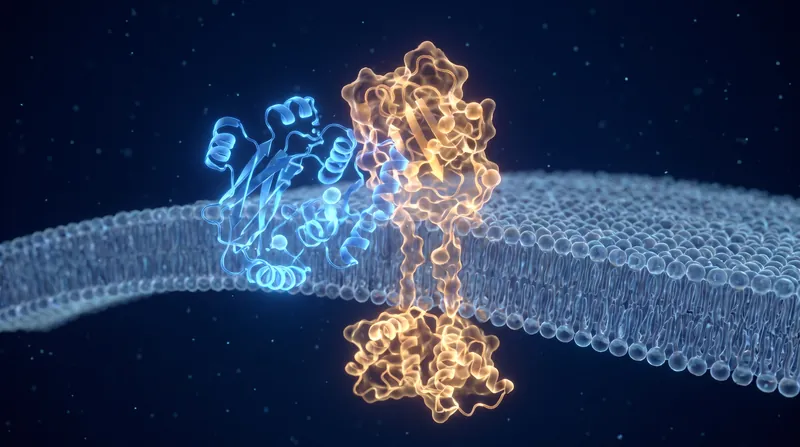
The vitamin D receptor (VDR) was first identified in 1969, fundamentally changing the scientific understanding of mineral metabolism. The VDR is a ligand-dependent transcription factor. When calcitriol binds to the VDR, it triggers a profound conformational change in the receptor's three-dimensional structure.[3][6]
This structural shift is the critical ignition key. However, the calcitriol-VDR complex cannot bind to DNA on its own. To become fully functional, it must find a partner. It heterodimerizes—or pairs up—with another nuclear receptor called the retinoid X receptor (RXR).[2][7]
However, the calcitriol-VDR complex cannot bind to DNA on its own.
The newly formed VDR-RXR heterodimer acts as a highly specific molecular scanner. It patrols the genome looking for exact sequences of DNA known as Vitamin D Response Elements (VDREs). These response elements are typically composed of two hexameric half-sites separated by three base pairs.[1][6]
When the heterodimer locates a VDRE, it docks onto the DNA strand. This physical attachment does not automatically transcribe the gene. Instead, the docked complex serves as a landing pad for a massive assembly of secondary proteins known as coactivators or corepressors.[2][6]
If the goal is to activate a gene, the complex recruits coactivator proteins with histone acetyltransferase (HAT) activity. These enzymes loosen the tightly wound chromatin, unwrapping the DNA spool and allowing the cell's transcription machinery to read the gene and produce messenger RNA.[1][3]
Conversely, if the hormone needs to turn a gene off, the complex recruits corepressors. These proteins bring histone deacetylases (HDACs) to the site, which tighten the chromatin structure, effectively locking the gene away and silencing its expression.[1][3]
The scale of this operation is vast. Genomic mapping detailed in the Journal of Clinical Investigation and the International Journal of Molecular Sciences reveals that the VDR regulates approximately 3 percent of the human genome. This translates to between 200 and 900 distinct genes, depending on the specific tissue and cellular environment.[1][2][7]
Historically, clinical focus remained on the classical targets: genes in the intestine, bone, and kidneys that manage calcium and phosphate absorption. This is why calcitriol is essential for preventing rickets and osteomalacia. The hormone upregulates calcium-binding proteins like calbindin to ensure mineral homeostasis.[4][5]
Modern genomic approaches have uncovered the hormone's non-classical reach. The VDR is expressed in nearly every tissue in the body, including activated T-cells, macrophages, and brain tissue. In these cells, calcitriol regulates genes involved in cellular proliferation, differentiation, and immune modulation.[2][4]
For the reader, this mechanism explains why maintaining adequate vitamin D levels is a systemic necessity rather than just a skeletal one. Because calcitriol operates at the foundational level of gene transcription, its effects are broad, powerful, and deeply integrated into the body's cellular defense and repair systems.[8]
It also explains why massive, sudden doses of vitamin D do not linearly increase biological benefit. The VDR can become saturated, and calcitriol strongly upregulates the CYP24A1 gene—which produces an enzyme that degrades calcitriol. This built-in negative feedback loop prevents toxicity but means that consistent, moderate levels are more effective for genomic regulation than erratic spikes.[1][5][8]
Jargon, explained
- Calcitriol
- The fully active, steroid hormone form of vitamin D, created after hydroxylation in the liver and kidneys.
- Vitamin D Receptor (VDR)
- A protein in the cell nucleus that binds to calcitriol and interacts with DNA to regulate gene expression.
- Retinoid X Receptor (RXR)
- A nuclear receptor that pairs with the VDR to form a complex capable of binding to specific DNA sequences.
- Transcription Factor
- A protein that controls the rate of transcription of genetic information from DNA to messenger RNA.
- Vitamin D Response Element (VDRE)
- Specific sequences of DNA that the VDR-RXR complex recognizes and binds to in order to control a target gene.
Sources
Source coverage
8 outlets
3 viewpoints surfaced






- [1]GenesGenomic Research
Vitamin D: An Overview of Gene Regulation, Ranging from Metabolism to Genomic Effects
Read on Genes → - [2]The Journal of Clinical InvestigationGenomic Research
The vitamin D receptor: contemporary genomic approaches reveal new basic and translational insights
Read on The Journal of Clinical Investigation → - [3]The Journal of Steroid Biochemistry and Molecular BiologyMolecular Biology
The Vitamin D Receptor Story: Discovery, Control, and Genomic Reach
Read on The Journal of Steroid Biochemistry and Molecular Biology → - [4]JBMR PlusMolecular Biology
Probing the Scope and Mechanisms of Calcitriol Actions Using Genetically Modified Mouse Models
Read on JBMR Plus → - [5]StatPearls (NCBI Bookshelf)Classical Endocrinology
Calcitriol
Read on StatPearls (NCBI Bookshelf) → - [6]Endocrinology and Metabolism Clinics of North AmericaClassical Endocrinology
The Vitamin D Receptor: New Paradigms for the Regulation of Gene Expression by 1,25-Dihydroxyvitamin D3
Read on Endocrinology and Metabolism Clinics of North America → - [7]International Journal of Molecular SciencesGenomic Research
Vitamin D and Its Receptor from a Structural Perspective
Read on International Journal of Molecular Sciences → - [8]Factlen Editorial TeamMolecular Biology
Synthesis by Factlen editorial team
Read on Factlen Editorial Team →
Comments

More in Health
See all →Metabolic Pathways
How Insulin Binding to the Tyrosine Kinase Receptor Activates the PI3K/Akt Pathway to Translocate GLUT4
10 sources
Drug Metabolism
The Specific Cytochrome P450 Isoforms That Metabolize 75% of Prescription Drugs
9 sources

Developmental Screening
The 75th Percentile Threshold: How the CDC Revised Developmental Milestones to Improve Early Screening
6 sources

Homeopathy Math
The Dilution Factor: Why Avogadro's Number Guarantees Zero Active Molecules in Most Homeopathic Remedies
5 sources

Every angle. Every day.
Get Health stories with full source coverage and perspective breakdowns delivered to your inbox.