Laser Phase Plate Breakthrough Solves an 80-Year Problem in Protein Imaging
Researchers have successfully integrated a 400-gigawatt laser into a cryo-electron microscope, dramatically boosting image contrast and making the cell's smallest, most elusive proteins visible for the first time.
By Factlen Editorial Team
- Structural Biologists
- Focus on the unprecedented ability to finally visualize the 90 percent of the proteome that was previously invisible to cryo-EM.
- Microscopy Engineers
- Emphasize the monumental technical achievement of stabilizing a 400-gigawatt laser cavity inside an electron microscope.
- Translational Researchers
- Highlight the downstream impacts for drug discovery and understanding the molecular basis of human disease.
What's not represented
- · Commercial microscope manufacturers
- · Crystallographers relying on traditional X-ray methods
Why this matters
For decades, more than 90 percent of the proteins inside human cells have been too small to image clearly, leaving scientists blind to the molecular roots of many diseases. By dramatically boosting microscope contrast, this breakthrough will allow researchers to see exactly how drugs interact with elusive proteins in their natural environment, accelerating the development of new therapeutics.
Key points
- Researchers have successfully integrated a laser phase plate into a cryo-electron microscope, solving an 80-year contrast problem.
- The technology uses a laser 100 million times brighter than the Sun to shift the phase of the microscope's electron beam.
- In paired experiments, the laser improved the resolution of small benchmark proteins like hemoglobin by up to 44 percent.
- A dual-laser system developed by CZ Biohub achieved a staggering 1.8 Ångstrom resolution on the apoferritin protein.
- The breakthrough could eventually allow scientists to image more than 50 percent of human proteins in their native cellular environments.
For decades, structural biologists have operated in the dark when it comes to the vast majority of the molecular machines that keep human cells alive.[7]
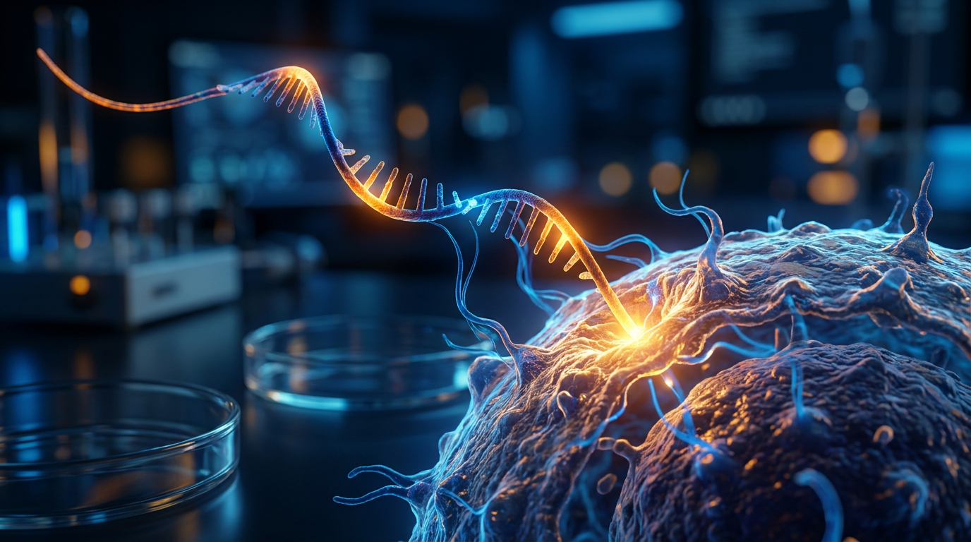
Cryo-electron microscopy (cryo-EM) revolutionized the field, earning a Nobel Prize by allowing scientists to flash-freeze proteins and image them at near-atomic resolution. The technology became the backbone of modern structural biology, accelerating drug discovery and revealing the architecture of viruses.[4][7]
Yet, a fundamental physics limitation remained: cryo-EM relies on electrons scattering off atoms to create an image, and small proteins simply do not scatter enough electrons to generate visible contrast. They appear washed out against the background noise.[5][6]
Because of this contrast problem, researchers estimate that more than 90 percent of the proteins found inside a human cell are too small for conventional cryo-EM to capture clearly, leaving them effectively invisible to science.[6]

Now, a landmark breakthrough published this week in Science and Nature Communications has provided a definitive solution to this 80-year-old imaging hurdle.[1][2]
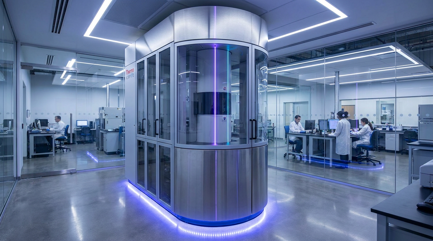
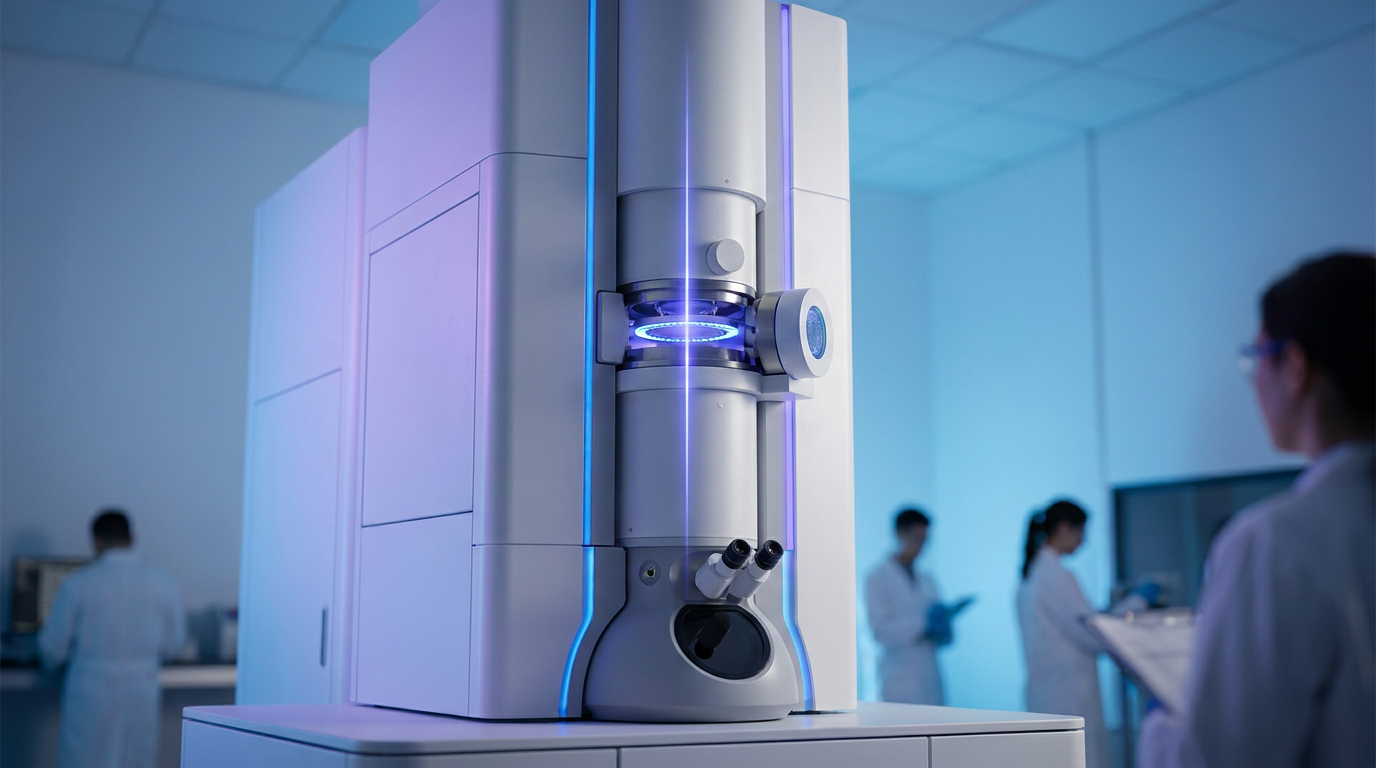
Teams from UC Berkeley and the Chan Zuckerberg (CZ) Biohub have successfully integrated a "laser phase plate" into a state-of-the-art cryo-electron microscope, a feat long dismissed by many in the field as practically impossible.[5][6]
The evidence pack presented across three coordinated publications demonstrates that this technology can dramatically sharpen images of small, elusive proteins, pushing the boundaries of what can be seen at the atomic level.[1][2][3]
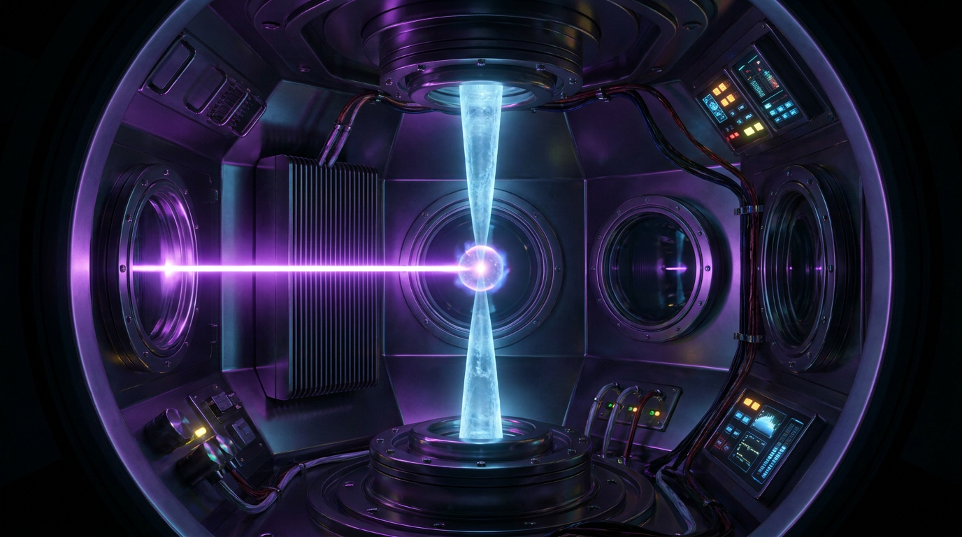
The mechanism relies on an extraordinary feat of optical engineering: bouncing a laser beam back and forth between two highly polished mirrors nearly 10,000 times inside a cavity the size of an espresso cup.[5][6]
This continuous bouncing creates a steady-state laser intensity of roughly 350 to 400 gigawatts per square centimeter—an energy density 100 million times brighter than the surface of the Sun, concentrated into a spot one-thousandth the width of a human hair.[6]

When the microscope's electron beam passes through this intense field of light, its phase is shifted. This optical manipulation translates into a massive boost in image contrast without destroying the delicate biological sample.[1][5]
When the microscope's electron beam passes through this intense field of light, its phase is shifted.
The empirical data is striking: in paired experiments, turning the laser on improved the resolution of hemoglobin—a benchmark small protein that sits at the very edge of conventional cryo-EM capabilities—by up to 44 percent.[1][5]
Features that were previously blurry and indistinct sharpened into clearly defined, high-resolution atomic structures, proving that the theoretical concept works flawlessly in practice.[1]

Furthermore, the CZ Biohub team introduced a second-generation "crossed laser phase plate" (xLPP), utilizing two perpendicular laser beams to split the power load and suppress optical artifacts known as ghost images.[2][3]
Using this dual-laser system, the researchers successfully imaged apoferritin, an iron-storage protein, at a staggering 1.8 Ångstroms, approaching the theoretical resolution limit of the technology.[3][6]
The implications extend far beyond imaging purified proteins in isolation; the true prize is cryo-electron tomography (cryo-ET), which images proteins in their native, crowded cellular environments.[4][6]
Currently, scientists can resolve fewer than 1 percent of proteins in their natural cellular context, but experts project the laser phase plate could make more than 50 percent of cellular proteins visible.[6]

This leap in visibility is expected to accelerate drug discovery, allowing researchers to watch exactly how a therapeutic molecule binds to a target protein amidst the chaotic jostling of a living cell.[6][7]
Despite the overwhelming success of the prototype, transparent uncertainties remain regarding the timeline for commercialization and widespread adoption across standard university laboratories.[7]
The current systems, including Berkeley's modified microscope dubbed "Theia," are bespoke, highly complex instruments that require demanding tolerances and specialized optical maintenance.[5][7]
Nevertheless, the successful demonstration of the laser phase plate marks a definitive turning point, transforming a theoretical concept into a practical tool that will illuminate the microscopic foundations of human health and disease.[7]
How we got here
1953
Frits Zernike wins the Nobel Prize in Physics for inventing the phase-contrast optical microscope.
2017
The Nobel Prize in Chemistry is awarded for the development of cryo-electron microscopy.
2010s
Physicists first propose using an intense laser to shift the phase of an electron beam, though many deem it practically impossible.
2021
CZ Biohub funds a major initiative to build a working prototype of the laser phase plate.
June 2026
Researchers publish coordinated papers demonstrating the successful integration and operation of single- and dual-laser phase plates in cryo-EM.
Viewpoints in depth
Structural Biologists
Focus on the unprecedented ability to finally visualize the 90 percent of the proteome that was previously invisible to cryo-EM.
For structural biologists, the laser phase plate represents the lifting of a decades-old fog. Because conventional cryo-EM relies on electrons scattering off atoms, small proteins simply do not generate enough signal to stand out against the background noise. This left researchers unable to study the vast majority of the molecular machines that drive cellular processes. By solving the contrast problem, biologists anticipate moving away from studying purified proteins in isolation and toward cryo-electron tomography (cryo-ET), which allows them to observe proteins interacting in their native, crowded cellular environments.
Microscopy Engineers
Emphasize the monumental technical achievement of stabilizing a 400-gigawatt laser cavity inside an electron microscope.
From an engineering perspective, the breakthrough is a triumph over extreme physical constraints. For years, the concept of a laser phase plate was considered theoretically elegant but practically impossible to build. Engineers had to figure out how to bounce a laser beam nearly 10,000 times between two mirrors inside a cavity the size of an espresso cup, generating an intensity 100 million times brighter than the Sun, all without destroying the delicate biological sample or the microscope itself. The introduction of the dual-laser crossed phase plate (xLPP) further solved issues of optical ghosting and power load distribution.
Translational Researchers
Highlight the downstream impacts for drug discovery and understanding the molecular basis of human disease.
Translational researchers view this technology as a massive accelerator for drug discovery. Currently, designing a drug often requires understanding the exact 3D shape of a target protein so a therapeutic molecule can be engineered to fit it perfectly. If a protein is too small to image, designing a drug for it involves significant guesswork. With the laser phase plate, researchers hope to clearly visualize these elusive targets and, crucially, watch how they bind to potential drugs in real-time within the cell, potentially unlocking treatments for diseases that have long evaded modern medicine.
What we don't know
- Whether the laser phase plate can be successfully miniaturized and commercialized for widespread use in standard university laboratories.
- The exact lower size limit of proteins that can be resolved; while hemoglobin (~64 kDa) was imaged, it remains unclear if proteins under 17 kDa will yield clear structures.
- How the intense laser cavity will perform over years of continuous operation, and what the long-term maintenance costs for the delicate mirror alignments will be.
Key terms
- Cryo-electron microscopy (cryo-EM)
- A technique that determines the atomic structure of molecules by bombarding flash-frozen samples with electrons.
- Phase contrast
- An optical technique that converts phase shifts in light (or electrons) passing through a transparent specimen into brightness changes in the image.
- Cryo-electron tomography (cryo-ET)
- An advanced imaging technique that captures multiple angular views of a sample to build a 3D model of proteins in their natural cellular environment.
- Ångstrom (Å)
- A unit of length equal to one ten-billionth of a meter, commonly used to express the size of atoms and molecules.
- Proteome
- The entire set of proteins that is, or can be, expressed by a genome, cell, tissue, or organism at a certain time.
Frequently asked
What is cryo-electron microscopy (cryo-EM)?
A Nobel Prize-winning technique that determines the 3D atomic structure of molecules by bombarding flash-frozen biological samples with electrons.
Why was a laser needed for the microscope?
Most proteins are too small to scatter enough electrons to create a clear image. The intense laser shifts the phase of the electron beam, dramatically boosting the image contrast.
Will this breakthrough cure diseases?
Not directly, but it allows scientists to see exactly how proteins interact and how drugs bind to them inside cells, which is a crucial step in designing better therapeutics.
Sources
Source coverage
7 outlets
3 viewpoints surfaced


[1]ScienceStructural Biologists
Enhanced contrast for small proteins with a single-laser phase plate in cryo-EM
Read on Science →[2]Nature CommunicationsMicroscopy Engineers
Theoretical framework for a dual-laser crossed phase plate in electron microscopy
Read on Nature Communications →[3]bioRxivStructural Biologists
Design and implementation of a crossed laser phase plate for cryo-electron microscopy
Read on bioRxiv →An innovative technology boosts image quality for protein structures
Read on Nature →[5]UC BerkeleyMicroscopy Engineers
A breakthrough in electron microscopy delivers sharper images of our body's tiniest proteins
Read on UC Berkeley →[6]CZ BiohubTranslational Researchers
Making the invisible visible: Microscope Breakthrough Will Open Unprecedented View into Our Cells
Read on CZ Biohub →[7]Factlen Editorial TeamTranslational Researchers
Synthesis by Factlen editorial team
Read on Factlen Editorial Team →

More in science
See all 74 stories →Cellular Imaging
The Laser Phase Plate: A Century-Old Physics Trick Just Unlocked the Human Proteome
5 sources
Cancer Vaccines
Personalized mRNA Cancer Vaccine Halves Melanoma Recurrence in 5-Year Milestone
8 sources
Cryo-EM Advance
Laser Phase Plate Breakthrough Illuminates the Smallest Proteins in Human Cells
7 sources
Ocean Tech
How Solar LEDs and Acoustic Pingers Are Solving the Global Bycatch Crisis
6 sources
Every angle. Every day.
Get science stories with full source coverage and perspective breakdowns delivered to your inbox.