Laser Phase Plate Breakthrough Allows Cryo-Electron Microscopes to Image the Body's Smallest Proteins
Researchers at UC Berkeley and Biohub have successfully integrated a laser phase plate into a cryo-electron microscope, solving an 80-year-old physics problem. The breakthrough dramatically boosts image contrast, allowing scientists to finally see the tiny proteins that make up 90% of the human body.
By Factlen Editorial Team
- Structural Biologists
- Focused on the immediate capability to resolve smaller proteins and map intact cells.
- Microscopy Engineers
- Focused on the technical achievement and the remaining thermal and stability hurdles.
- Computational Biologists
- Focused on generating high-fidelity ground truth data for AI models.
What's not represented
- · Pharmaceutical Companies
- · Commercial Microscope Manufacturers
Why this matters
More than 90% of the proteins in our bodies are too small to be seen clearly with current microscopes, leaving scientists blind to the exact mechanisms of many diseases. By using a laser to dramatically boost image contrast, this breakthrough allows researchers to finally see these elusive proteins in their natural cellular environment, paving the way for highly targeted new drugs.
Key points
- Researchers have successfully integrated a laser phase plate into a cryo-electron microscope, solving an 80-year-old physics problem.
- The device uses a laser 100 million times brighter than the Sun to shift the electron beam's phase, dramatically boosting image contrast.
- Conventional cryo-EM is blind to roughly 90% of human proteins because they are too small to generate sufficient contrast.
- The breakthrough allowed scientists to clearly image hemoglobin, a benchmark small protein at the lower limit of older machines.
- The technology promises to revolutionize cryo-electron tomography, allowing scientists to map proteins operating inside intact cells.
- Engineers are already developing a dual-beam 'crossed' laser system to reduce thermal stress and eliminate ghost images.
For decades, structural biologists have been peering into the fundamental machinery of life through a glass, darkly. Cryo-electron microscopy (cryo-EM) revolutionized our understanding of biology by revealing the atomic architecture of proteins, earning a Nobel Prize in 2017. Yet, the technique harbors a massive blind spot: it relies on electrons scattering off molecules to create an image, and small proteins simply do not scatter enough electrons to be seen clearly against the background noise.[3][4]
This physical limitation means that conventional cryo-EM is effectively blind to roughly 90 percent of the proteins found inside human cells. To image them, scientists must extract them from their native environment, purify them, and study them in isolation—a process that strips away the crucial context of how these molecular machines interact, assemble, and malfunction in living tissue.[4][5]
On June 11, 2026, a collaborative team from UC Berkeley and Biohub published a landmark breakthrough in the journal Science (and highlighted in Nature) that promises to shatter this barrier. After more than fifteen years of theoretical proposals and engineering dead-ends, the researchers successfully integrated a "laser phase plate" (LPP) into a state-of-the-art cryo-electron microscope, dramatically boosting the contrast of the images it produces.[1][2][3]

The concept of phase contrast is not new; it was introduced to optical microscopy nearly a century ago by Frits Zernike, who won the 1953 Nobel Prize in Physics for the discovery. Zernike realized that transparent biological cells shift the phase of light passing through them. By using a physical plate to shift the unscattered background light by 90 degrees, he converted invisible phase differences into stark, high-contrast amplitude differences.[1][3]
Translating Zernike’s optical trick to electron microscopy, however, has proven notoriously difficult. Electrons are charged particles. When researchers tried inserting physical phase plates—typically ultra-thin carbon films—into the electron beam, the plates quickly accumulated electrostatic charge. This stray charge distorted the electron wave, scrambling the image and rendering the technique practically useless for high-resolution structural biology.[1]
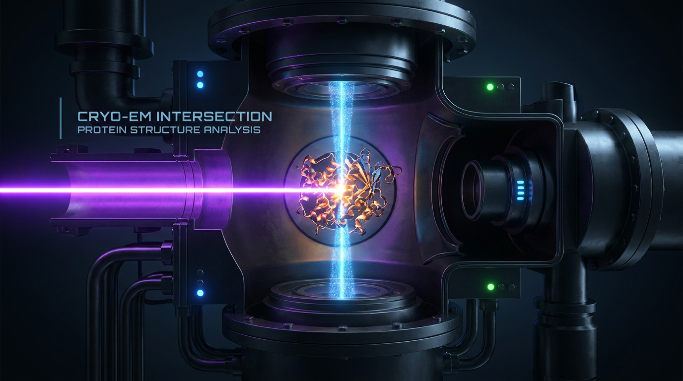
The UC Berkeley and Biohub teams bypassed the charging problem entirely by replacing the physical plate with pure light. Their system utilizes a continuous-wave laser, trapped and amplified between two highly polished mirrors inside the microscope's vacuum chamber. As the laser bounces back and forth, it builds to an intensity roughly 100 million times brighter than the surface of the Sun.[1][4]
When the microscope's electron beam passes through this intense optical field, the electromagnetic energy of the laser shifts the phase of the electrons by exactly 90 degrees via a phenomenon known as the ponderomotive effect. Because the electrons interact only with photons—not with a physical material—there is zero electrostatic charging, zero unwanted scattering, and zero degradation of the phase plate over time.[1][7]
The evidence for the LPP’s efficacy is robust. In their Science publication, the UC Berkeley team, led by physicist Holger Müller, demonstrated the system's power by imaging hemoglobin. Hemoglobin, the protein responsible for carrying oxygen in red blood cells, is relatively small and sits at the absolute lower size limit for conventional cryo-EM machines.[1][3]

In their Science publication, the UC Berkeley team, led by physicist Holger Müller, demonstrated the system's power by imaging hemoglobin.
The resulting images show a stark improvement. The laser phase plate significantly enhanced the contrast and resolution of the hemoglobin structure compared to standard imaging techniques. For structural biologists, this is the equivalent of turning on the headlights in a dark room; proteins that were previously washed out in the background noise suddenly snap into sharp focus.[1][3]
But the true prize of this technology extends far beyond imaging isolated proteins. The LPP is poised to revolutionize a newer, more complex technique known as cryo-electron tomography (cryo-ET). While single-particle cryo-EM takes thousands of pictures of purified proteins to build a 3D model, cryo-ET tilts an intact, flash-frozen cell to capture multiple angular views, reconstructing a 3D volume of the cell's interior.[1][2][4]
Currently, the contrast in cryo-ET is so poor that researchers estimate fewer than 1 percent of proteins can be reliably identified in their native cellular environment. Biohub scientists project that the laser phase plate could push that number past 50 percent. This would allow researchers to map the crowded, dynamic interior of cells at near-atomic resolution, observing exactly how drugs bind to targets or how viruses hijack cellular machinery in real time.[4][5]
Despite the breakthrough, the technology remains in its infancy, and significant engineering hurdles remain before it becomes a standard fixture in structural biology core facilities. Operating a laser at 100 million times solar intensity inside a highly sensitive electron microscope introduces massive thermal management challenges. The cavity mirrors must maintain sub-nanometer stability while absorbing intense heat, a problem the current papers acknowledge requires ongoing refinement.[5]

Furthermore, the intense single-beam laser can create "ghost images"—faint, duplicate copies of high-contrast objects that can obscure the delicate biological signals researchers are trying to isolate. To address this, Biohub simultaneously released a preprint describing a second-generation "crossed laser phase plate" (xLPP).[5][6]
The xLPP design splits the laser power between two intersecting beams arranged in an X-shape. This dual-beam approach not only suppresses the ghosting artifact but also halves the intensity required from each individual mirror cavity, reducing the thermal stress on the system and making the instrument significantly more stable for routine use.[5][6]
We view the laser phase plate not just as a better microscope, but as a critical data engine for the future of biology. As artificial intelligence models like AlphaFold increasingly dominate protein structure prediction, they require massive amounts of ground-truth data to validate their in-cell predictions. The LPP provides the hardware necessary to make the interior of the human cell machine-readable at an unprecedented scale.[5][7]
How we got here
1953
Frits Zernike wins the Nobel Prize in Physics for inventing the phase-contrast optical microscope.
2017
The Nobel Prize in Chemistry is awarded for the development of cryo-electron microscopy (cryo-EM).
2010s
The 'resolution revolution' in cryo-EM allows atomic-level imaging of large purified proteins, accelerating drug discovery.
June 11, 2026
UC Berkeley and Biohub researchers publish the successful integration of a laser phase plate into a cryo-EM system in Science.
Viewpoints in depth
Structural Biologists
Focused on the immediate capability to resolve smaller proteins and map intact cells.
For researchers studying the molecular basis of disease, the laser phase plate is a paradigm shift. By extending cryo-EM's reach to the 90% of the human proteome previously obscured by low contrast, biologists can finally image elusive drug targets. The ultimate goal is to transition from studying purified proteins in isolation to observing them dynamically interacting within the crowded environment of a living cell via cryo-electron tomography.
Microscopy Engineers
Focused on the technical achievement and the remaining thermal and stability hurdles.
Engineers view the LPP as the elegant solution to an 80-year-old physics problem: how to achieve phase contrast in an electron microscope without the electrostatic charging that ruins physical plates. However, they emphasize that maintaining a continuous-wave laser at 100 million times solar intensity inside a vacuum chamber requires extreme thermal management. The development of the dual-beam crossed laser phase plate (xLPP) is seen as the necessary next step to reduce thermal stress and eliminate optical artifacts.
Computational Biologists
Focused on generating high-fidelity ground truth data for AI models.
As AI models like AlphaFold revolutionize protein structure prediction, their next frontier is predicting how complexes assemble and function inside the cell. Computational biologists see the LPP not just as a microscope, but as a data engine. By dramatically increasing the volume of high-resolution, in-cell structural data, the LPP will provide the essential ground truth needed to train and validate the next generation of predictive biological AI.
What we don't know
- How quickly the laser phase plate can be commercialized and scaled to structural biology core facilities worldwide.
- Whether the intense thermal output of the laser cavity can be managed reliably enough for high-throughput, 24/7 automated data collection.
- How effectively the second-generation 'crossed' laser design will suppress ghost images in highly crowded cellular samples.
Key terms
- Cryo-electron microscopy (cryo-EM)
- A technique that fires a beam of electrons at flash-frozen biological samples to determine the 3D atomic structure of molecules.
- Phase contrast
- An imaging technique that converts invisible shifts in a wave's phase (caused by passing through a transparent object) into visible changes in brightness.
- Ponderomotive effect
- A phenomenon where charged particles, like electrons, are pushed or have their phase shifted by an intense, oscillating electromagnetic field, such as a laser.
- Proteome
- The entire set of proteins that is, or can be, expressed by a genome, cell, tissue, or organism at a certain time.
- Cryo-electron tomography (cryo-ET)
- An advanced imaging technique that tilts a frozen sample to capture multiple views, creating a 3D reconstruction of an intact cell's interior.
Frequently asked
What is cryo-electron microscopy (cryo-EM)?
Cryo-EM is a Nobel-winning technique that fires electrons at flash-frozen biological samples to determine the atomic structure of proteins and molecules.
Why couldn't we see small proteins before?
Small proteins don't scatter enough electrons to create a clear image against the background noise, making them practically invisible to conventional cryo-EM.
How does the laser phase plate fix this?
It uses an intensely focused laser to shift the phase of the electron beam by 90 degrees, converting invisible phase differences into high-contrast visible images without physically touching the beam.
What is cryo-electron tomography (cryo-ET)?
Cryo-ET is an advanced technique that takes multiple angled images of an intact cell to build a 3D model of its interior, allowing scientists to see proteins operating in their natural environment.
Sources
Source coverage
7 outlets
3 viewpoints surfaced


[1]ScienceStructural Biologists
Laser phase plate improves structure determination of small proteins by cryo-EM
Read on Science →[2]NatureStructural Biologists
An innovative technology boosts image quality for protein structures
Read on Nature →[3]UC BerkeleyStructural Biologists
A breakthrough in electron microscopy delivers sharper images of our body's tiniest proteins
Read on UC Berkeley →[4]BiohubMicroscopy Engineers
Microscope Breakthrough Will Open Unprecedented View into Our Cells
Read on Biohub →[5]LabcriticsComputational Biologists
Laser Phase Plate Becomes Reality: Biohub and Berkeley Solve Cryo-EM's 80-Year Contrast Problem with a Laser
Read on Labcritics →[6]bioRxivMicroscopy Engineers
A dual-beam crossed laser phase plate for high-contrast cryo-electron microscopy
Read on bioRxiv →[7]Factlen Editorial TeamComputational Biologists
Synthesis by Factlen editorial team
Read on Factlen Editorial Team →
Every angle. Every day.
Get science stories with full source coverage and perspective breakdowns delivered to your inbox.