An Innovative Laser Technology Dramatically Boosts Image Quality for Protein Structures
Researchers have successfully integrated a high-intensity laser into an electron microscope, solving a decades-old physics problem. The breakthrough allows scientists to image the smallest proteins inside human cells, opening a new frontier in structural biology and drug discovery.
By Factlen Editorial Team
- Structural Biologists
- Focused on mapping the fundamental architecture of life and seeing proteins interact in their native cellular environment.
- Microscopy Engineers
- Focused on overcoming the physical limits of electron optics and integrating high-power lasers into vacuum chambers.
- Drug Discovery Researchers
- Focused on translating high-resolution structural insights into targeted therapeutics for complex diseases.
What's not represented
- · Commercial microscope manufacturers who must figure out how to mass-produce the technology.
- · Computational biologists who must design new software to process the novel phase-contrast data.
Why this matters
More than 90 percent of human proteins are currently too small to be seen clearly by standard electron microscopes. By making these invisible molecular machines visible, this technology could unlock new treatments for diseases that currently lack druggable targets.
Key points
- Conventional cryo-electron microscopy struggles to image small proteins, leaving 90% of human proteins difficult to resolve.
- Researchers have successfully integrated a high-intensity laser into a cryo-EM to act as a 'phase plate'.
- The laser shifts the phase of the electron beam, dramatically increasing image contrast without destroying high-resolution data.
- The breakthrough could eventually allow scientists to see how proteins interact inside intact, living cells.
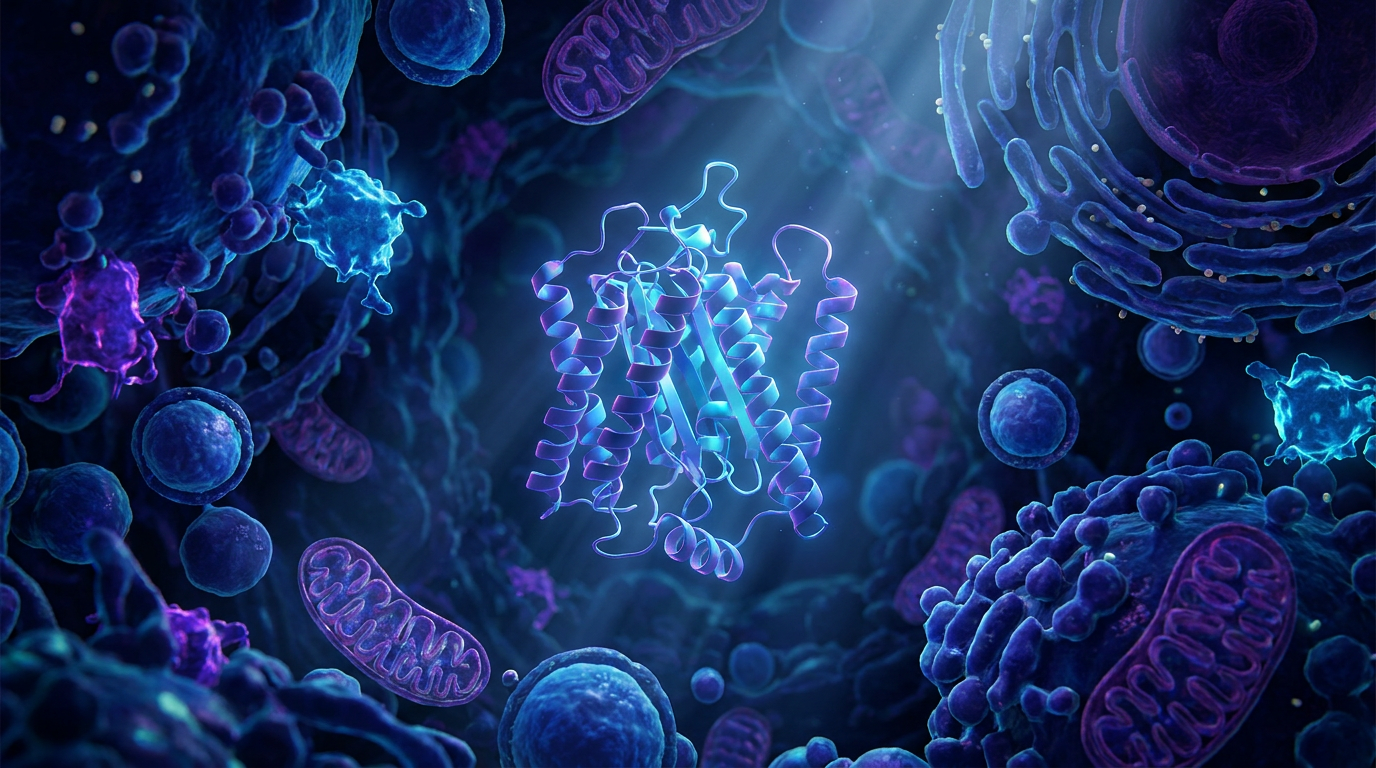
Inside every human cell, tens of thousands of proteins act as the molecular machinery of life—ferrying cargo, repairing DNA, and triggering cellular division. For decades, structural biologists have sought to map these machines atom by atom. The advent of cryo-electron microscopy (cryo-EM) revolutionized this quest, earning a Nobel Prize in 2017 for allowing scientists to visualize purified proteins frozen in vitreous ice. Yet, despite its immense power, conventional cryo-EM has hit a physical wall. It struggles to generate enough contrast to clearly image small molecules, leaving a massive blind spot in our understanding of human biology.[1][4][5]
That blind spot is now shrinking. In a landmark achievement published in Science and highlighted by Nature, researchers from UC Berkeley and Biohub have successfully integrated a "laser phase plate" into a state-of-the-art electron microscope. The device uses a continuous-wave laser, amplified to an intensity 100 million times brighter than the surface of the sun, to manipulate the electron beam. This optical intervention dramatically boosts the contrast of the resulting images, allowing scientists to resolve proteins that were previously too small or faint to see.[2][3][4][5][6][7]
The scale of the problem this solves is difficult to overstate. More than 90 percent of the proteins found inside human cells are simply too small for conventional cryo-EM to capture clearly. Currently, scientists estimate that only about 10 percent of the human proteome can be imaged in purified form, and fewer than 1 percent can be visualized in their native, crowded cellular environments. By fundamentally altering how the microscope detects biological matter, the laser phase plate promises to push that boundary, potentially making more than 50 percent of cellular proteins visible to researchers.[5]

To understand the breakthrough, one must understand the "phase problem" inherent to electron microscopy. When an electron beam passes through a biological specimen, the sample does not absorb the electrons to create a dark shadow. Biological molecules are composed of light elements like carbon, oxygen, and nitrogen, which act as "weak phase objects." Instead of blocking the electrons, the protein merely slows them down slightly, shifting the phase of their quantum wave function.[1][2][7]
Because standard detectors only measure the amplitude (intensity) of electrons, this subtle phase shift is virtually invisible. For decades, microscopists bypassed this problem by deliberately throwing the microscope out of focus. Defocusing the lens converts some of the phase shift into detectable amplitude contrast, making the protein visible. However, this optical trick comes at a steep cost: it destroys high-resolution data, blurring the finest atomic details of the molecule.[2][6][7]
The ideal solution is a physical phase plate—a device placed in the path of the electron beam that shifts the phase of the unscattered electrons by exactly 90 degrees. This perfectly converts the specimen's phase modulation into sharp amplitude contrast without requiring the lens to be defocused. Nearly a century ago, a similar concept called phase-contrast optical microscopy won a Nobel Prize for making transparent cells visible under light microscopes.[2][4][7]
Translating that concept to electron microscopes, however, proved maddeningly difficult. Previous attempts relied on thin carbon films to shift the electron phase. But when a powerful electron beam strikes a physical material, it rapidly deposits an electrostatic charge. These carbon phase plates would charge up and degrade almost instantly, rendering them unstable and largely impractical for routine, high-resolution data collection.[7]
Translating that concept to electron microscopes, however, proved maddeningly difficult.
The solution, first proposed more than 15 years ago by UC Berkeley physicist Holger Müller and biophysicist Robert Glaeser, was to replace the physical carbon film with light. Because photons do not carry an electrical charge, a laser beam cannot suffer from electrostatic charging. However, electrons and photons barely interact. To force a phase shift, the laser would need to be unimaginably intense.[5][7]
The newly unveiled system achieves this by firing a continuous-wave laser into an optical cavity consisting of highly polished, precision mirrors. The mirrors bounce the light back and forth, building a standing wave of immense power. When the electron beam passes through the focused focal point of this laser, it encounters a phenomenon known as the ponderomotive potential. The intense electromagnetic field of the laser slightly retards the electrons, shifting their phase by the crucial 90 degrees without scattering them.[4][5][6][7]

Building the device required years of engineering to overcome immense technical hurdles. A proof-of-concept was demonstrated in 2019 on an older generation microscope, but integrating the delicate optical cavity into a modern, state-of-the-art Thermo Scientific Krios cryo-EM machine required a massive collaborative effort between UC Berkeley and Biohub. The resulting instrument is one of the most complex microscopes ever constructed.[5][7]
To prove the system's efficacy, the research teams tested it on biological benchmarks. They first imaged aldolase, a medium-sized enzyme that is relatively easy for conventional machines to resolve. The laser phase plate delivered a clear improvement in resolution. But the true test was hemoglobin, the oxygen-carrying protein in red blood cells.[4][6]
At a molecular weight of just 64 kilodaltons, hemoglobin sits at the absolute lower frontier of what conventional cryo-EM can detect. It is right at the edge of the field's vision. When imaged with the new laser phase plate, the contrast of the hemoglobin molecules was dramatically enhanced, allowing the software to assemble a highly detailed 3D structure. The researchers noted that the smaller the sample, the greater the relative advantage provided by the laser.[4][5][6]

While imaging purified proteins is a massive step forward, the ultimate prize is cryo-electron tomography (cryo-ET). Cryo-ET involves tilting a flash-frozen cell and taking multiple images from different angles to compute a 3D reconstruction of the intact cellular environment. This allows scientists to see proteins not in isolation, but interacting with their neighbors inside the cell.[4][5]
Because cells are thick and complex, cryo-ET suffers from notoriously low contrast. The laser phase plate is uniquely suited to solve this. By providing in-focus phase contrast, the technology could allow researchers to map the interior of cells with unprecedented clarity, creating sub-nanometer atlases of health and disease. "To see all those interactions has been the dream of structural cell biologists for decades," noted David Agard, Biohub's Founding Scientific Director of Imaging.[2][5][6]

Despite the triumph, the technology remains in its infancy, and several uncertainties lie ahead. Single-beam laser designs can produce "ghost images"—faint, artifactual copies of high-contrast objects that can obscure delicate biological signals. Biohub is actively developing a dual-laser variant to suppress these artifacts and distribute the thermal load on the mirrors. Furthermore, scaling this bespoke, highly customized optical setup into a commercial product that can be deployed to laboratories worldwide will require significant time and industrial investment.[1][5]
Nevertheless, the successful demonstration of the laser phase plate marks a watershed moment in structural biology. By making the invisible visible, the technology promises to accelerate drug discovery, revealing the hidden molecular targets that drive cancer, neurodegeneration, and infectious diseases. As the field transitions from looking at isolated parts to observing the entire cellular engine in motion, the laser phase plate may well be the lens that brings the future of medicine into focus.[1][4][5]
How we got here
1953
Phase-contrast optical microscopy wins the Nobel Prize in Physics.
2010
Holger Müller and Robert Glaeser first propose the concept of a laser phase plate for electron microscopes.
2017
Cryo-electron microscopy (cryo-EM) wins the Nobel Prize in Chemistry.
2019
The first proof-of-concept of a laser phase plate is demonstrated on an older generation microscope.
June 2026
UC Berkeley and Biohub publish the successful integration of the laser phase plate into a state-of-the-art cryo-EM.
Viewpoints in depth
Structural Biologists
Focused on mapping the fundamental architecture of life and seeing proteins in their native environment.
For structural biologists, the laser phase plate represents the holy grail of cellular imaging. For decades, the field has been forced to study proteins in isolation—purifying them in a test tube and freezing them in ice. While this has yielded incredible insights into protein shapes, it strips away the biological context. Biologists argue that to truly understand how a cell functions, they must see proteins interacting with their neighbors in the crowded, dynamic environment of the intact cell. By providing the contrast necessary for high-resolution cryo-electron tomography (cryo-ET), this technology finally makes that vision possible.
Microscopy Engineers
Focused on overcoming the physical limits of electron optics and integrating high-power lasers into vacuum chambers.
From an engineering perspective, the laser phase plate is a triumph over seemingly insurmountable physical constraints. Previous attempts to create phase plates using physical materials like carbon films failed because the electron beam rapidly deposited an electrostatic charge, ruining the image. Using light instead of matter solves the charging problem, but introduces immense new challenges: aligning a continuous-wave laser, amplified to 100 million times the brightness of the sun, inside the delicate vacuum chamber of an electron microscope. Engineers are now focused on refining dual-laser designs to eliminate optical artifacts like 'ghost images' and making the system robust enough for commercial production.
Drug Discovery Researchers
Focused on translating high-resolution structural insights into targeted therapeutics.
The pharmaceutical industry relies heavily on structural biology to design drugs that fit perfectly into the active sites of disease-causing proteins. However, because conventional cryo-EM struggles to resolve small proteins, many critical targets in cancer, neurodegeneration, and infectious diseases have remained 'undruggable.' Drug discovery researchers view the laser phase plate as a vital tool to unlock these targets. By dramatically improving the resolution of small molecules, the technology will allow chemists to see exactly how potential drug compounds bind to their targets, accelerating the development of more effective and precise medicines.
What we don't know
- How quickly the bespoke laser phase plate technology can be commercialized and scaled for widespread laboratory use.
- Whether the dual-laser design will fully eliminate the 'ghost image' artifacts observed in the single-beam prototypes.
- The exact upper limit of resolution the system can achieve when applied to the most complex, crowded cellular environments in cryo-ET.
Key terms
- Cryo-electron microscopy (cryo-EM)
- A technique that fires electrons at flash-frozen biological samples to determine their 3D atomic structure.
- Phase contrast
- An optical technique that converts invisible phase shifts in a wave (caused by passing through transparent matter) into visible changes in brightness.
- Ponderomotive potential
- The physical force exerted by an intense, oscillating electromagnetic field (like a laser) that can shift the phase of passing electrons.
- Cryo-electron tomography (cryo-ET)
- A 3D imaging technique that takes multiple pictures of a tilted, intact cell to show proteins in their natural environment.
- Kilodalton (kDa)
- A unit of mass used in biochemistry to measure the size of proteins and other large molecules.
Frequently asked
Why can't scientists just use regular light microscopes to see proteins?
Proteins are much smaller than the wavelength of visible light, making them physically impossible to resolve with optical microscopes. Electron microscopes use electron beams, which have much shorter wavelengths, to see atomic details.
How does a laser affect an electron beam if photons have no charge?
While photons don't have an electrical charge, an incredibly intense laser creates a strong electromagnetic field. This field exerts a 'ponderomotive potential' that slightly slows down the electrons, shifting their phase.
When will this technology be used to discover new drugs?
While the prototype is currently operational in research settings, scaling the technology into commercial microscopes that pharmaceutical companies can buy will likely take several years of industrial development.
Sources
Source coverage
7 outlets
3 viewpoints surfaced


[1]Factlen Editorial TeamDrug Discovery Researchers
Synthesis by Factlen editorial team
Read on Factlen Editorial Team →[2]NatureStructural Biologists
An innovative technology boosts image quality for protein structures
Read on Nature →[3]ScienceMicroscopy Engineers
Laser phase plate improves structure determination of small proteins by cryo-EM
Read on Science →[4]UC BerkeleyMicroscopy Engineers
Physicists introduce phase contrast to electron microscopy, delivering sharper images of our body's tiniest proteins
Read on UC Berkeley →[5]BiohubStructural Biologists
Making the invisible visible – laser phase plate cryo-EM
Read on Biohub →[6]News-MedicalDrug Discovery Researchers
Laser phase plate dramatically expands cryo-EM imaging capabilities
Read on News-Medical →[7]National Institutes of HealthMicroscopy Engineers
Laser phase plate for transmission electron microscopy
Read on National Institutes of Health →
Every angle. Every day.
Get science stories with full source coverage and perspective breakdowns delivered to your inbox.