A 15-Year Quest to Build a 'Laser Phase Plate' Just Unlocked the Invisible Human Proteome
By integrating a laser 100 million times brighter than the sun into an electron microscope, physicists have finally solved cryo-EM's biggest limitation, allowing scientists to see the atomic structures of the body's smallest proteins.
By Factlen Editorial Team
- Structural Biologists
- Eager to map the 'dark matter' of the human proteome and unlock structures of small proteins.
- Cryo-EM Physicists
- View the LPP as a triumph over a 15-year theoretical engineering challenge.
- Industry Analysts & Synthesis
- Anticipate that higher-resolution structures of elusive targets will accelerate drug design and commercialization.
What's not represented
- · Commercial Microscope Manufacturers
- · AI Protein Folding Researchers
Why this matters
More than 90 percent of human proteins are too small to be seen by conventional electron microscopes. This breakthrough allows researchers to finally visualize these elusive molecules, dramatically accelerating the discovery of new drugs for diseases ranging from cancer to Alzheimer's.
Key points
- A new 'laser phase plate' dramatically improves the contrast of cryo-electron microscopes.
- The technology allows scientists to image proteins smaller than 70 kilodaltons for the first time.
- Roughly 90 percent of human proteins fall into this previously invisible size category.
- The device uses a continuous-wave laser 100 million times brighter than the sun.
- Researchers successfully demonstrated the tech by imaging hemoglobin at high resolution.
- The breakthrough is expected to significantly accelerate structure-based drug discovery.
For decades, biologists have been trying to map the intricate molecular machinery that keeps human cells alive. But they have been operating with a massive blind spot.[6]
The revolutionary technique known as cryo-electron microscopy (cryo-EM) earned a Nobel Prize in 2017 for allowing scientists to see the atomic structure of biological molecules. By flash-freezing samples and bombarding them with electrons, researchers could finally visualize the complex proteins that drive life.[1][3]
Yet, cryo-EM has a fundamental physical limitation: it struggles to see anything small. Proteins below a mass of roughly 70 kilodaltons simply do not scatter enough electrons to stand out against the background of frozen water.[3][4]
This is not a niche problem. Approximately 90 percent of the proteins in the human body—the very molecules responsible for countless diseases and the targets for most drugs—fall below this 70-kilodalton threshold. They are effectively invisible to the world's most powerful microscopes.[3][4][5]

Now, a breakthrough 15 years in the making promises to turn the lights on. In a series of papers published this week in Science and Nature Communications, researchers from UC Berkeley and the Chan Zuckerberg Biohub unveiled a working "laser phase plate" (LPP).[2][3][5]
The LPP solves the contrast problem by borrowing a trick from optical microscopy. Nearly a century ago, scientists discovered that shifting the phase of light waves as they pass through a transparent sample creates visible contrast—a discovery that also won a Nobel Prize.[3]
Applying this phase-contrast principle to an electron beam, however, was long considered practically impossible. It requires interfering with a beam of electrons traveling at a significant fraction of the speed of light without destroying the delicate biological sample.[4][5]
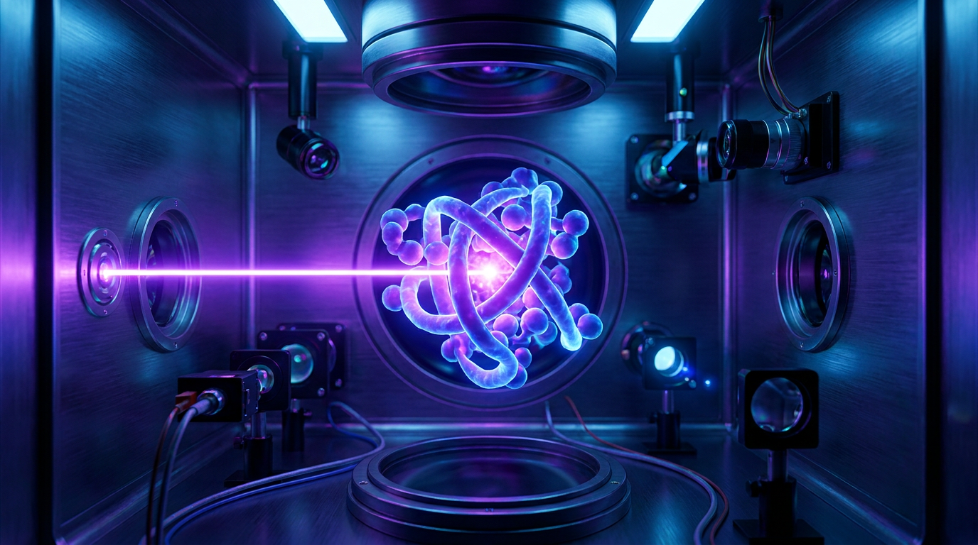
Physicists Holger Müller and Robert Glaeser first proposed the theoretical solution more than a decade ago: use a laser. By trapping a continuous-wave laser inside a tiny mirrored cavity, they could create a wall of light intense enough to slightly slow down the passing electrons.[3][4]
Physicists Holger Müller and Robert Glaeser first proposed the theoretical solution more than a decade ago: use a laser.
The engineering required to build this was staggering. The resulting laser cavity, installed inside a custom Thermo Fisher Krios microscope dubbed "Theia," generates a beam 100 million times more intense than the surface of the sun, concentrated into a space of a few microns.[4][5]

When the electron beam passes through this intense optical field, its phase shifts. The result is a dramatic, immediate boost in the signal-to-noise ratio of the final image.[1][2]
To prove the concept, the Berkeley and Biohub teams imaged hemoglobin, the oxygen-carrying protein in blood. At roughly 64 kilodaltons, hemoglobin sits right at the ragged edge of what conventional cryo-EM can resolve, often appearing as a blurry blob.[3][5]
With the laser phase plate engaged, the blurry blob sharpened into a clearly defined, high-resolution atomic structure. The LPP improved resolution by up to 44 percent in paired experiments, proving that the theoretical physics translated into tangible biological insight.[3][5]
The implications extend far beyond isolated proteins. The LPP is expected to supercharge a related technique called cryo-electron tomography (cryo-ET), which takes multiple angular views of a sample to build 3D models of intact cells.[1][3]
Currently, finding a specific small protein inside the crowded, chaotic environment of a whole cell using cryo-ET is like "trying to find one leaf on one tree in a forest," as one Biohub director described it. The LPP provides the contrast needed to pick that leaf out from the background noise.[3]

For the pharmaceutical industry, this represents a paradigm shift. If researchers can clearly see the atomic structure of the small proteins involved in cancer, Alzheimer's, or autoimmune diseases, they can design drugs that bind to them with unprecedented precision.[2][4]
The challenge now shifts from fundamental physics to commercial engineering. The current LPP systems are bespoke, highly sensitive prototypes running in specialized physics labs.[5][6]
Both the Berkeley and Biohub teams are collaborating with major microscope manufacturers to miniaturize and stabilize the technology. The goal is to make the laser phase plate a standard, push-button upgrade for the hundreds of cryo-EM facilities operating worldwide.[5]
How we got here
1953
Frits Zernike wins the Nobel Prize for inventing phase-contrast light microscopy.
2010s
Advances in direct electron detectors trigger the 'resolution revolution' in cryo-EM.
2011
Physicists Holger Müller and Robert Glaeser first propose using a laser to shift the phase of an electron beam.
2021
The Chan Zuckerberg Biohub provides major funding to build a working prototype of the laser phase plate.
June 2026
Researchers publish successful demonstrations of the LPP imaging small proteins in Science and Nature Communications.
Viewpoints in depth
Structural Biologists
Eager to map the 'dark matter' of the human proteome.
For structural biologists, the laser phase plate is the equivalent of launching the James Webb Space Telescope. They have spent the last decade mapping the large, easily visible macromolecular complexes of the cell, but have been frustrated by the inability to see the smaller proteins that do much of the actual work. By breaking the 70-kilodalton barrier, they can finally begin cataloging the structures of the remaining 90 percent of the proteome, shifting their focus from what the microscope allows them to see to what biology actually demands they study.
Cryo-EM Physicists
Focused on pushing the theoretical limits of electron optics.
The engineering community views the LPP as a triumph over a problem that many considered practically impossible. Interfering with an electron beam using light requires a laser cavity of staggering intensity, perfectly aligned and stabilized to within fractions of a nanometer. Their success proves that manipulating electron phases with continuous-wave lasers is not just a theoretical curiosity, but a viable path forward for the next generation of atomic-resolution imaging.
Pharmaceutical Researchers
Looking to leverage high-resolution structures for targeted drug design.
Drug discovery relies heavily on structure-based design—if you know the exact 3D shape of a disease-causing protein, you can design a chemical key that fits perfectly into its lock. Because the vast majority of drug targets are small proteins that conventional cryo-EM cannot resolve, pharmaceutical researchers have had to rely on less accurate predictive models. The LPP promises to provide empirical, high-resolution maps of these elusive targets, potentially shaving years off the drug development pipeline and leading to more effective, highly specific medications.
What we don't know
- How quickly the bespoke laser phase plate technology can be miniaturized and commercialized for standard laboratory use.
- Whether the intense laser cavity will introduce unforeseen artifacts when imaging highly sensitive or unstable protein complexes.
- The exact lower size limit the technology will ultimately reach, though researchers are targeting proteins as small as 17 kilodaltons.
Key terms
- Cryo-Electron Microscopy (Cryo-EM)
- A technique that fires electrons at flash-frozen biological samples to determine their 3D atomic structure.
- Proteome
- The entire set of proteins that is, or can be, expressed by a genome, cell, tissue, or organism.
- Kilodalton (kDa)
- A unit of mass used by biologists to measure the size of proteins and other large molecules.
- Phase Contrast
- A method of converting slight shifts in how a wave passes through a transparent object into visible differences in brightness.
- Cryo-Electron Tomography (Cryo-ET)
- An advanced technique that takes multiple 2D images of a sample from different angles to build a 3D model of an intact cell.
Frequently asked
Why couldn't we just use regular microscopes to see these proteins?
Light microscopes are limited by the wavelength of visible light, which is too large to resolve individual atoms. Electron microscopes use much shorter wavelengths, allowing for atomic-level resolution.
How does the laser actually help the electron microscope?
The intense laser light acts as a 'lens' that slightly slows down the electron beam, shifting its phase. When these shifted electrons hit the detector, they create a much higher contrast image of the nearly transparent protein.
When will this technology be available for drug discovery?
While the prototype is currently working in specialized physics labs, it will likely take several years to commercialize and integrate the laser phase plate into standard cryo-EM machines used by pharmaceutical companies.
Sources
Source coverage
6 outlets
3 viewpoints surfaced


[1]NatureStructural Biologists
An innovative technology boosts image quality for protein structures
Read on Nature →[2]ScienceStructural Biologists
Laser-boosted microscopes could reveal new drug targets, sharpen views inside cells
Read on Science →[3]UC BerkeleyCryo-EM Physicists
A breakthrough in electron microscopy delivers sharper images of our body's tiniest proteins
Read on UC Berkeley →[4]BiohubCryo-EM Physicists
Microscope Breakthrough Will Open Unprecedented View into Our Cells
Read on Biohub →[5]LabCriticsIndustry Analysts & Synthesis
The Laser Phase Plate is Real
Read on LabCritics →[6]Factlen Editorial TeamIndustry Analysts & Synthesis
Synthesis by Factlen editorial team
Read on Factlen Editorial Team →
Every angle. Every day.
Get science stories with full source coverage and perspective breakdowns delivered to your inbox.