New AI Models Detect Pancreatic Cancer Years Before Human Doctors Can
An AI system developed by the Mayo Clinic can identify microscopic signs of pancreatic cancer on routine CT scans up to three years before conventional diagnosis, doubling the detection rate of expert radiologists.
By Factlen Editorial Team
- Clinical Researchers
- Focused on the life-saving potential of identifying cancer before it forms a visible tumor.
- Open-Source Advocates
- Pushing to democratize medical AI so that global health systems can build upon foundational models.
- Medical Practitioners
- Cautiously optimistic, prioritizing safe workflow integration and avoiding false-positive fatigue.
What's not represented
- · Health Insurance Providers
- · Patient Advocacy Groups
Why this matters
Pancreatic cancer is one of the most lethal malignancies precisely because it grows silently and is usually caught too late. By leveraging AI to find microscopic tissue changes years in advance, doctors could shift the disease from a near-certain death sentence to a highly treatable condition.
Key points
- The Mayo Clinic's REDMOD AI can detect pancreatic cancer on routine CT scans up to three years before a conventional diagnosis.
- In retrospective studies, the AI achieved 73% sensitivity, compared to 39% for expert human radiologists reviewing the same scans.
- The system detects 'imaging-occult' cancer by analyzing microscopic textural disruptions in the tissue before a tumor is visible.
- Alibaba's DAMO Academy has developed similar breakthrough AI models for early pancreatic and stomach cancer detection.
- Open-source initiatives are simultaneously releasing foundational medical AI models to democratize advanced diagnostics globally.
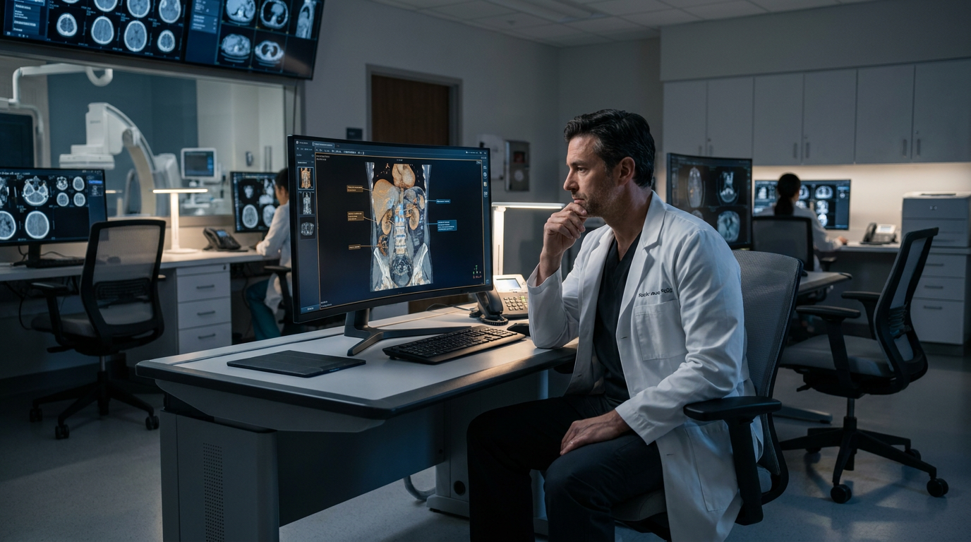
Pancreatic cancer has long been one of medicine's most elusive and lethal killers, largely because it grows silently and is rarely detected before it has spread to other organs. Now, a wave of artificial intelligence breakthroughs is fundamentally changing that timeline, offering a vital new window for early intervention. A newly developed AI system from the Mayo Clinic, known as REDMOD, can spot the disease on routine abdominal CT scans up to three years before a conventional diagnosis would typically occur.[2]
Published recently in the British Society of Gastroenterology's medical journal Gut, the Mayo Clinic study revealed that REDMOD specifically targets what researchers classify as 'imaging-occult' cancer. These are highly elusive cases where no visible tumor exists—even upon rigorous expert re-review of the scans—yet the disease is already biologically underway. The cancer is encoded in microscopic tissue changes and cellular remodeling that no human eye, regardless of the radiologist's experience or the monitor's resolution, can naturally detect during a standard examination.[1][2]
In a rigorous retrospective test against expert human radiologists, REDMOD correctly identified nearly three in four pre-diagnostic scans, achieving a remarkable 73 percent sensitivity rate. Human radiologists reviewing the exact same scans caught less than 39 percent of the early-stage cases. The performance gap became even more pronounced at longer time horizons; at lead times stretching beyond two years before a clinical diagnosis, the artificial intelligence was nearly three times more sensitive than its highly trained human counterparts.[2][5]

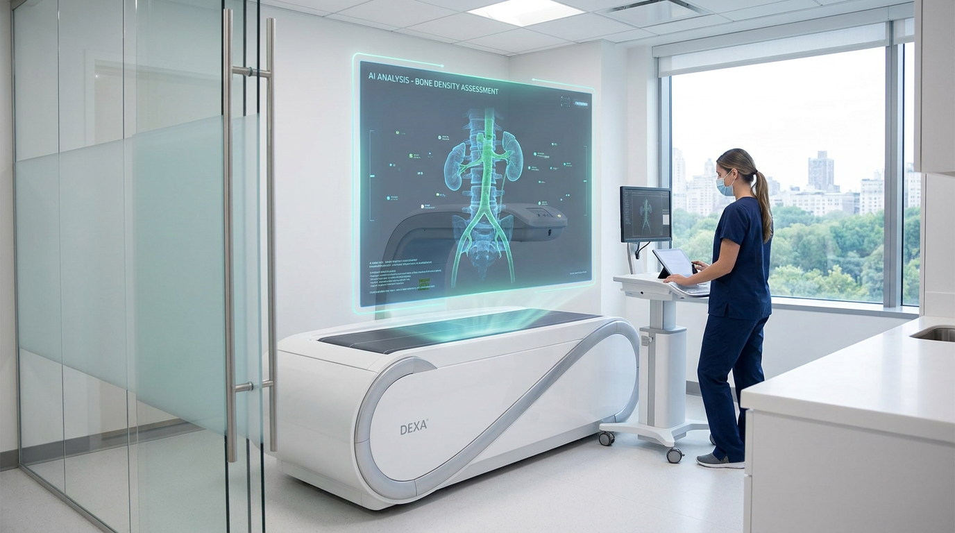
The system achieves this unprecedented accuracy by analyzing nearly 1,000 distinct radiomic features extracted from the pancreas, eventually narrowing them down to 40 key predictive signals. The vast majority of these critical signals are derived from wavelet-filtered images—a sophisticated mathematical technique that exposes subtle textural disruptions in the tissue. Researchers believe these microscopic disruptions reflect the early biological remodeling of the organ, serving as a digital footprint of the cancer long before a physical tumor actually forms.[1][2]
The Mayo Clinic's success is part of a broader, global surge in AI-driven oncology that is rapidly moving from the laboratory to the clinic. In China, researchers at Zhejiang Cancer Hospital and Alibaba's DAMO Academy previously developed PANDA, an AI model also designed to detect pancreatic cancer through ordinary non-contrast CT imaging. PANDA demonstrated a staggering 92.9 percent sensitivity in clinical trials and was officially granted breakthrough device designation by the U.S. Food and Drug Administration in April 2025.[3]
The Mayo Clinic's success is part of a broader, global surge in AI-driven oncology that is rapidly moving from the laboratory to the clinic.
Building on that regulatory and clinical success, the same Chinese research coalition recently unveiled GRAPE, a specialized model capable of detecting early-stage stomach cancer. Deployed in multiple hospitals across the Zhejiang and Anhui provinces, GRAPE boosted overall detection rates by up to 24.5 percent in real-world trials. Notably, the system identified asymptomatic stomach cancer in roughly 40 percent of its detected cases, proving that image-based gastric screening is highly viable even without the use of specialized contrast dyes.[3][4]

While proprietary models like REDMOD and PANDA push the absolute boundaries of clinical accuracy, a parallel movement is working aggressively to democratize these diagnostic tools. Eindhoven University of Technology recently unveiled a massive open-source AI model trained on over 250,000 CT scans. By making the foundational architecture freely available to the global medical community, the university hopes hospitals worldwide can tailor the AI to their specific demographic needs without paying exorbitant licensing fees to private technology companies.[7]
Major technology companies are also fueling this open-source healthcare ecosystem, recognizing that collaborative development accelerates innovation. Meta's open-source Llama foundation models are increasingly being adapted by medical startups like Zauron Labs to double-check radiological imaging for human errors. Similarly, European biotechnology startups are building universal AI foundation models specifically for pathology, aiming to identify genetic abnormalities in tissue samples at a fraction of the traditional cost, thereby lowering the barrier to entry for advanced medical research.[6][8]

The next major hurdle for the medical community is integrating these hyper-sensitive AI tools into daily hospital workflows without overwhelming doctors. REDMOD features an adjustable detection threshold, allowing clinicians to carefully balance diagnostic sensitivity against the very real risk of false positives. Its baseline precision already exceeds the 3 percent threshold set by the UK's National Institute for Health and Care Excellence for acceptable cancer referrals, ensuring that the system flags genuine risks without causing unnecessary patient anxiety.[2]
Because these AI models analyze routine CT scans that patients already undergo for entirely unrelated issues—such as kidney stones, appendicitis, or abdominal pain—they require no new invasive procedures. For high-risk groups, such as patients with sudden-onset diabetes or a family history of the disease, this opportunistic screening could soon become a standard background check. The AI will quietly scan for microscopic threats, potentially shifting pancreatic cancer from a near-certain death sentence to a highly treatable condition.[1][2]
How we got here
April 2025
Alibaba's PANDA AI model for pancreatic cancer receives FDA breakthrough device designation.
July 2025
Chinese researchers publish data on GRAPE, an AI model capable of detecting early-stage stomach cancer from standard CT scans.
November 2025
Eindhoven University of Technology (TU/e) releases a massive open-source AI model for medical imaging analysis.
May 2026
The Mayo Clinic publishes research on REDMOD, demonstrating its ability to detect pancreatic cancer up to three years early.
Viewpoints in depth
Clinical Researchers
Focused on the life-saving potential of identifying cancer before it forms a visible tumor.
For oncologists and researchers at institutions like the Mayo Clinic and Zhejiang Cancer Hospital, the primary value of these AI models is shifting the timeline of intervention. Pancreatic and stomach cancers are notoriously lethal because they are almost always caught in advanced stages. By identifying 'imaging-occult' signatures—microscopic tissue remodeling that precedes a physical tumor—doctors can intervene when the disease is most treatable. They view opportunistic screening of routine CT scans as a low-cost, high-reward strategy that could drastically reduce mortality rates without requiring new, invasive testing protocols.
Open-Source Advocates
Pushing to democratize medical AI so that global health systems can build upon foundational models.
Computer scientists and open-source proponents argue that proprietary, locked-down medical AI models will only exacerbate global health inequalities. Institutions like Eindhoven University of Technology advocate for releasing foundational models trained on massive datasets to the public. They argue that by providing the 'stem' of the AI, hospitals in developing nations or underfunded research centers can fine-tune the models for their specific populations. This camp believes that the sheer volume of medical data and potential applications is too vast for any single company to monopolize, making open collaboration essential.
Medical Practitioners
Cautiously optimistic, prioritizing safe workflow integration and avoiding false-positive fatigue.
Frontline radiologists and hospital administrators welcome the diagnostic assistance but remain highly focused on the practicalities of deployment. Their primary concern is 'alarm fatigue'—if an AI system is too sensitive, it could flag thousands of healthy patients for unnecessary, anxiety-inducing follow-up biopsies. Practitioners emphasize that AI must act as a 'second set of eyes' rather than an autonomous decision-maker. They strongly support models with adjustable detection thresholds, allowing human doctors to calibrate the AI's sensitivity based on a patient's individual risk factors, such as a recent diabetes diagnosis or family history.
What we don't know
- How the AI models will perform across highly diverse, global patient populations outside of their initial training datasets.
- The exact clinical protocols hospitals will adopt for patients flagged by the AI who do not yet show any physical tumors.
- Whether insurance companies will cover the cost of preventative treatments based solely on an AI's radiomic risk assessment.
Key terms
- Radiomics
- The extraction of a large number of quantitative features from medical images using data-characterization algorithms, uncovering disease characteristics invisible to the naked eye.
- Imaging-occult cancer
- Cancer that is present biologically but cannot be seen on standard medical imaging, even by expert human reviewers.
- Wavelet-filtered images
- A mathematical technique used in image processing to separate data into different frequency components, helping to reveal subtle textural patterns in tissue.
- Opportunistic screening
- The practice of using medical tests or imaging conducted for one reason to simultaneously check for other, unrelated diseases.
- Sensitivity
- In medical diagnostics, the ability of a test to correctly identify patients who actually have the disease (true positive rate).
Frequently asked
Does this mean I need to get a special CT scan?
No. The AI models are designed to analyze standard, routine CT scans that patients might undergo for entirely unrelated reasons, such as abdominal pain or kidney stones.
Will the AI replace human radiologists?
No. The AI is designed to augment human doctors by flagging microscopic tissue changes. A human radiologist and oncologist will always review the findings and make the final diagnostic and treatment decisions.
Is this technology available in all hospitals right now?
Not yet. While some models like PANDA and GRAPE are deployed in select hospitals, tools like REDMOD are still undergoing clinical workflow integration and regulatory validation before widespread adoption.
Why is early detection so important for pancreatic cancer?
Pancreatic cancer rarely shows symptoms in its early stages. By the time a patient feels sick, the cancer has usually spread to other organs, making it incredibly difficult to treat.
Sources
Source coverage
8 outlets
3 viewpoints surfaced


[1]GutClinical Researchers
Identification of occult radiomic signatures for early pancreatic cancer detection using REDMOD
Read on Gut →[2]Mayo Clinic PressClinical Researchers
AI Breakthrough Detects Pancreatic Cancer Up to Three Years Before Diagnosis
Read on Mayo Clinic Press →[3]XinhuaClinical Researchers
China's AI breakthrough set to be game-changer for early cancer detection
Read on Xinhua →[4]Nature MedicineClinical Researchers
Non-contrast CT imaging for early gastric cancer detection via GRAPE AI
Read on Nature Medicine →[5]AI Breakthrough TrackerClinical Researchers
How effective is Mayo Clinic's AI for pancreatic cancer detection?
Read on AI Breakthrough Tracker →[6]VivaTechMedical Practitioners
The AI Checkup: Diagnosing the Future of Healthcare
Read on VivaTech →[7]Eindhoven University of TechnologyOpen-Source Advocates
TU/e unveils open-source AI model to enhance medical diagnosis
Read on Eindhoven University of Technology →[8]MetaOpen-Source Advocates
How Open Source AI is Evolving Healthcare
Read on Meta →

Every angle. Every day.
Get ai stories with full source coverage and perspective breakdowns delivered to your inbox.