AI Repurposes Routine Bone Scans to Detect Hidden Heart Disease Years Before Symptoms
A new AI algorithm can analyze standard bone density scans to spot early signs of cardiovascular disease in seconds, potentially turning millions of routine osteoporosis checks into life-saving heart screenings.
By Factlen Editorial Team
- Clinical Researchers
- Focuses on the technical achievement of training algorithms to spot calcification faster and more accurately than human specialists.
- Public Health Advocates
- Emphasizes the cost-efficiency of repurposing existing medical infrastructure to screen millions without new equipment.
- Preventative Care Patients
- Highlights the psychological shift from silent risk to actionable data, empowering individuals to make lifestyle changes.
What's not represented
- · Radiology Technicians
- · Health Insurance Providers
Why this matters
Heart disease is the leading cause of death globally, often remaining 'silent' until a catastrophic event occurs. By piggybacking on the millions of DEXA bone scans already performed annually, this AI tool offers a zero-cost, zero-radiation method to catch arterial calcification years before a heart attack strikes.
Key points
- A new AI algorithm detects abdominal aortic calcification (AAC) using standard DEXA bone density scans.
- AAC is a major early warning sign of cardiovascular disease that often appears before heart blockages.
- The AI analyzes the scans in seconds, replacing a slow and expensive manual review by specialists.
- The technology could passively screen millions of patients annually without additional costs or radiation exposure.
Heart disease remains the leading cause of death worldwide, yet it frequently operates in the shadows. For millions of people, the first noticeable symptom of cardiovascular trouble is a catastrophic event, such as a heart attack or stroke. Alarmingly, roughly one in five heart attacks are considered "silent," meaning the damage occurs before the patient even realizes they are at risk. Traditional risk assessments, which rely heavily on indirect indicators like cholesterol levels and blood pressure, often fail to provide a complete picture of a patient's actual arterial health. Now, a major breakthrough in artificial intelligence is poised to change that dynamic, offering a way to peer inside the body's blood vessels long before a crisis strikes.[1]
An international team of researchers, led by Professor Joshua Lewis at Edith Cowan University in Western Australia, has successfully trained an artificial intelligence algorithm to detect early markers of heart disease using routine bone scans. The breakthrough demonstrates that AI can accurately identify dangerous calcium build-ups in the body's largest artery. By repurposing existing medical imaging, the technology offers a cost-effective, highly scalable method for preventative screening.[2][3]
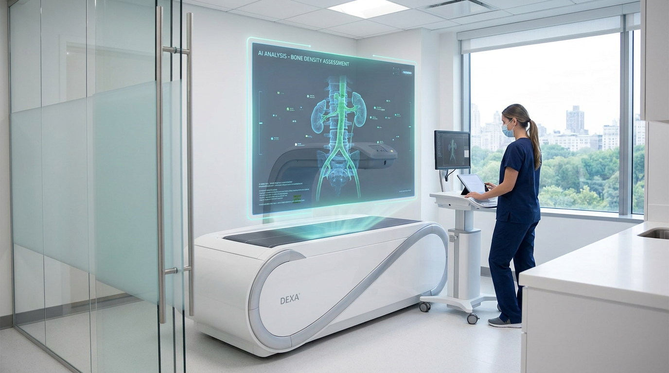
The innovation centers on dual-energy x-ray absorptiometry, commonly known as DEXA scans. These low-dose x-ray machines are a staple of modern medicine, primarily used to assess bone mineral density and diagnose osteoporosis in older adults. Because the scans capture clear lateral images of the spine and surrounding tissues, they inadvertently photograph the abdominal aorta. For years, researchers have known that these images contained valuable cardiovascular data, but extracting that information was a logistical challenge.[1][5]
The specific marker the AI looks for is abdominal aortic calcification, or AAC. This condition involves the gradual accumulation of calcium deposits within the walls of the aorta. Medical professionals recognize AAC as a powerful, independent predictor of advanced blood vessel disease and future cardiovascular events. Crucially, this calcification often appears in the abdominal aorta years before similar blockages develop in the arteries directly supplying the heart.[1]

Catching AAC early provides a critical window for intervention, but doing so has historically been difficult. Detecting the calcification on a standard DEXA scan requires a highly trained specialist to manually review the lateral spine images. This process is painstakingly slow, expensive, and generally cost-prohibitive for widespread population screening. As a result, the cardiovascular data captured during millions of routine bone scans has largely been ignored, leaving patients unaware of their hidden risks.[3][4]
The newly developed AI algorithm eliminates this bottleneck entirely. The software automatically analyzes the lateral spine images captured by the DEXA machine, detecting and quantifying the exact level of abdominal aortic calcification. What once required a specialist's time-consuming review is now accomplished by the algorithm in a matter of seconds. The system is designed to complement existing diagnostic workflows, delivering fast, reliable results without requiring any additional effort from the radiology technician.[1][2]
The newly developed AI algorithm eliminates this bottleneck entirely.
The public health implications of this technology are massive, largely because it piggybacks on existing medical infrastructure. In Australia alone, approximately 700,000 people undergo DEXA bone density testing every year. In the United States, that number exceeds three million. By integrating the AI software into the machines already sitting in hospitals and clinics worldwide, healthcare systems can instantly transform a single-purpose osteoporosis check into a dual-diagnostic tool, screening millions for heart disease at zero additional cost and with zero extra radiation exposure.[1][5]
The project has garnered significant support from the medical community, including backing from the Heart Foundation's Catalyst Partnership program. Professor Garry Jennings, the Heart Foundation's Chief Medical Advisor, praised the discovery as a vital development in preventative cardiology. The research team is now actively seeking further backing from private and philanthropic funders to accelerate the software's regulatory approval and bring the intervention to clinical settings as quickly as possible.[1][3]

For patients, the transition from invisible risk to actionable data can be life-altering. Pippa Luke, a patient who participated in the research, discovered she had advanced blood vessel disease only after the AI analyzed her scan. She noted that without the technology, her condition would have remained entirely silent. The stark results served as an immediate wake-up call, prompting her to take control of her cardiovascular health before the calcification could trigger a heart attack.[3][4]
Armed with the knowledge of her AAC risk, Luke fundamentally overhauled her lifestyle. She adopted a Mediterranean diet, incorporated regular cardiovascular and resistance training into her routine, and began scheduling frequent heart health checks with her physician. Clinical data shows that providing patients with their AAC results can drive measurable improvements in cardiovascular risk factors within just twelve weeks, proving that early awareness directly translates into better health outcomes.[1][3][4]
This breakthrough represents a broader paradigm shift in how artificial intelligence is being deployed in cardiovascular medicine. Beyond bone scans, researchers are increasingly using AI to extract hidden data from other routine tests, such as measuring pericardial fat from standard CT scans or quantifying arterial calcification in mammograms. By training algorithms to see what the human eye might miss—or what human specialists simply do not have the time to look for—technology is closing the gap between medical discovery and everyday care delivery.[2][6]
Professor Lewis, who first began exploring the link between DEXA scans and heart disease in 2009 at Sir Charles Gairdner Hospital, views the AI as an empowering tool rather than a replacement for doctors. The goal is to provide clinicians with better, faster data so they can intervene earlier. Whether through dietary changes, exercise, or preventative medications, giving doctors a multi-year head start on heart disease dramatically increases the odds of patient survival.[2][4]

The research team is currently focused on the next phase of deployment, which involves securing the necessary regulatory approvals to roll the software out to commercial radiology clinics. Because the AI utilizes machines that are already universally available, the barrier to entry is significantly lower than it would be for a completely new diagnostic device. Researchers hope to see the technology available in everyday clinical practice within the next few years.[3][4]
Ultimately, the DEXA scan breakthrough highlights the immense potential of looking at old data through a new lens. By applying cutting-edge artificial intelligence to routine, decades-old imaging techniques, medical science has unlocked a powerful new defense against the world's deadliest disease. It is a testament to the idea that the next great leap in healthcare might not require inventing a new machine, but simply teaching the machines we already have to look a little closer.[1][2]
How we got here
2009
Professor Joshua Lewis begins exploring the link between DEXA bone scans and heart disease at Sir Charles Gairdner Hospital.
2025
The Heart Foundation awards funding to the project through its Catalyst Partnership program to develop the AI algorithm.
May 2026
The research team publishes breakthrough findings demonstrating the AI's ability to accurately detect calcification in seconds.
Mid-2026
Researchers begin seeking regulatory approval to deploy the AI software in everyday clinical settings.
Viewpoints in depth
Clinical Researchers
The technical triumph of dual-diagnostic imaging.
For medical researchers, the DEXA scan breakthrough is a proof-of-concept for the future of radiology. The challenge was never a lack of data; the abdominal aorta has always been visible in lateral spine x-rays. The hurdle was the human bottleneck. By training an algorithm to instantly quantify abdominal aortic calcification, researchers have proven that single-purpose medical machines can be transformed into multi-diagnostic tools via software updates alone, bypassing the need to invent or manufacture new hardware.
Public Health Advocates
Scaling preventative care without scaling costs.
Public health organizations view this AI application as a massive win for health equity and preventative medicine. Cardiovascular disease screening often requires dedicated appointments, bloodwork, and specialized imaging, which creates friction for patients. By piggybacking on the millions of bone density scans already performed annually for osteoporosis, health systems can passively screen a massive swath of the aging population for heart disease at zero additional cost and with zero extra radiation exposure.
Preventative Care Patients
Transforming invisible threats into actionable lifestyle changes.
From the patient perspective, the value of the AI lies in its ability to make the invisible visible. Because early-stage arterial calcification produces no symptoms, patients often feel perfectly healthy right up until they suffer a heart attack. Providing patients with concrete, personalized data about their blood vessel health serves as a powerful psychological catalyst. Clinical trials show that patients who see their AAC results are significantly more likely to adopt Mediterranean diets, increase their exercise, and adhere to preventative medications.
What we don't know
- Exactly how long the regulatory approval process will take before the AI is available in standard commercial radiology clinics.
- Whether health insurance providers will automatically cover the dual-diagnostic AI analysis as part of a routine bone density check.
Key terms
- Abdominal Aortic Calcification (AAC)
- The build-up of calcium deposits in the walls of the abdominal aorta, serving as a strong predictor of future cardiovascular events.
- DEXA Scan
- A specialized, low-dose X-ray test used to measure bone mineral density, typically to check for osteoporosis.
- Cardiovascular Disease
- A broad term for conditions affecting the heart or blood vessels, often associated with a build-up of fatty deposits inside the arteries.
- Asymptomatic Risk
- A medical condition or risk factor that does not show any outward symptoms, making it difficult to detect without screening.
Frequently asked
What is a DEXA scan?
A dual-energy x-ray absorptiometry (DEXA) scan is a low-dose x-ray primarily used to measure bone mineral density and diagnose osteoporosis.
What does the AI look for?
The AI analyzes the scans for abdominal aortic calcification (AAC), which is a build-up of calcium in the body's largest artery and an early warning sign of heart disease.
Does this require an extra medical appointment?
No. The AI analyzes the exact same images already taken during a routine bone density check, meaning no extra time or radiation for the patient.
When will this be available to the public?
Researchers are currently seeking regulatory approval to deploy the AI software in clinical settings, hoping to roll it out within the next few years.
Sources
Source coverage
6 outlets
3 viewpoints surfaced


[1]Heart FoundationPublic Health Advocates
AI detects heart disease using DEXA scans
Read on Heart Foundation →[2]Edith Cowan UniversityClinical Researchers
Medical research meets AI
Read on Edith Cowan University →[3]The West AustralianPreventative Care Patients
Major West Australian AI breakthrough finds heart disease can be detected early in common bone scans
Read on The West Australian →[4]Sir Charles Gairdner HospitalClinical Researchers
AI breakthrough in heart disease began at Charlies
Read on Sir Charles Gairdner Hospital →[5]Mirage NewsPreventative Care Patients
AI Detects Heart Disease Using DEXA Bone Scans
Read on Mirage News →[6]News-MedicalPublic Health Advocates
Measuring heart fat with AI improves cardiovascular risk prediction
Read on News-Medical →

More in ai
See all 5 stories →


Every angle. Every day.
Get ai stories with full source coverage and perspective breakdowns delivered to your inbox.