Laser Phase Plate Breakthrough Solves 80-Year Contrast Problem in Electron Microscopy
A new laser device boosts the contrast of cryo-electron microscopes, allowing scientists to clearly image the small proteins that make up 90 percent of the human body.
By Factlen Editorial Team
- Structural Biologists
- Focused on the immediate gains in resolution and the ability to map previously invisible drug targets.
- Bioengineering Innovators
- Focused on the mechanical achievement of the device and the open-science approach to its development.
- Technology Evaluators
- Focused on the gap between proof-of-principle experiments and the ultimate goal of in-cell tomography.
What's not represented
- · Pharmaceutical Companies
- · AI Algorithm Developers
Why this matters
By allowing scientists to clearly see the tiny proteins that make up 90 percent of the human body, this breakthrough will dramatically accelerate the discovery of new drugs and treatments for diseases that were previously impossible to study at the molecular level.
Key points
- A new laser phase plate solves an 80-year-old physics problem, dramatically boosting the contrast of cryo-electron microscopes.
- The technology allows scientists to image proteins as small as 50 kilodaltons, unlocking access to 90 percent of the human proteome.
- Peer-reviewed evidence shows the laser improved the resolution of the hemoglobin protein by up to 44 percent.
- The device uses a continuous-wave laser 100 million times brighter than the sun to shift the phase of the electron beam.
- While proven on purified proteins, researchers are now working to apply the technology to intact cells using cryo-electron tomography.
For decades, structural biologists have been mapping the molecular machinery of life, but their most powerful tool has suffered from a profound blind spot. Cryo-electron microscopy (cryo-EM), a technique that won the Nobel Prize in 2017, fires beams of electrons at flash-frozen samples to reveal the intricate 3D shapes of biological molecules. Yet, despite its revolutionary impact on drug discovery and disease research, the technology is fundamentally limited by a physics problem: it cannot clearly see small things. Now, an engineering breakthrough published in Nature and Science has demonstrated a functional laser phase plate, a device that dramatically boosts the contrast of electron microscopes. By focusing a laser 100 million times brighter than the sun onto the electron beam, researchers from UC Berkeley and the Chan Zuckerberg Biohub have successfully imaged proteins previously considered too small to resolve.[1][2][3][5]
The primary claim advanced by the research teams is that this technology solves an 80-year-old contrast problem that has kept the vast majority of human biology in the dark. To understand the stakes, one must look at the scale of the limitation. Conventional cryo-EM struggles to produce clear images of proteins smaller than roughly 70 kilodaltons. Because small proteins do not scatter enough electrons to stand out from the background noise, they appear as faint, uninterpretable blobs. According to Biohub researchers, this means that approximately 90 percent of the human proteome—including countless critical disease mechanisms and potential drug targets—is effectively invisible to current cryo-EM techniques.[3][4][5][6]
The theoretical solution to this problem has been known since 1953, when Dutch physicist Frits Zernike won a Nobel Prize for introducing phase contrast to light microscopy. Zernike discovered that shifting the phase of light waves as they pass through a transparent sample converts invisible optical delays into stark, visible contrast. For more than 15 years, physicists Holger Müller and Robert Glaeser of UC Berkeley theorized that the exact same principle could be applied to electron microscopes. However, the engineering required to manipulate a high-energy electron beam without destroying the microscope was widely dismissed by the field as practically impossible.[2][3][4][6]

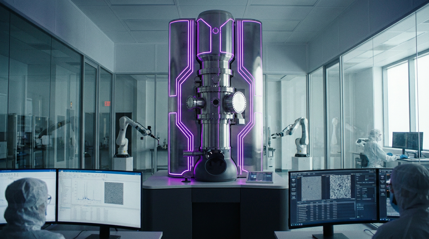
The newly published evidence demonstrates that the impossible has been engineered into reality. The UC Berkeley team successfully installed a customized optical cavity inside a 14-foot-tall Thermo Fisher Krios electron microscope. Inside this cavity, highly polished mirrors amplify a continuous-wave laser to 75 kilowatts of power, focusing it down to a width of just a few microns. When the electron beam passes through this intense field of photons, its phase is shifted, artificially boosting the signal-to-noise ratio of the resulting image. The researchers claim this hardware will eventually allow biologists to image proteins as small as 17 kilodaltons, unlocking nearly the entire human proteome.[2][3][5]
The strongest empirical evidence for the phase plate's efficacy comes from paired laser-on and laser-off experiments detailed in the foundational Science publication. To benchmark the device, the researchers imaged hemoglobin, a 64-kilodalton blood protein that sits at the absolute lower frontier of what conventional cryo-EM can resolve. The results provided robust, peer-reviewed validation of the underlying physics: activating the laser improved the resolution of the hemoglobin structure by up to 44 percent. Features that were previously lost in the background noise sharpened into clearly defined, high-contrast molecular architectures.[2][4]
While the single-laser system proved the concept, a parallel preprint released by Biohub engineers offers evidence for how the technology will scale to commercial viability. A single 75-kilowatt laser places immense physical stress on the microscope's internal mirrors, creating a persistent risk of catastrophic hardware failure. To mitigate this, the Biohub team designed a second-generation crossed laser phase plate (xLPP) that utilizes two intersecting laser beams. This dual-beam approach splits the power load, reducing the strain on the components while simultaneously suppressing the faint ghost images that can sometimes obscure the primary biological signal.[4][5]
A single 75-kilowatt laser places immense physical stress on the microscope's internal mirrors, creating a persistent risk of catastrophic hardware failure.
Despite these impressive engineering milestones, the evidence pack surrounding the technology's ultimate application remains incomplete. The stated long-term goal of the laser phase plate is not merely to image purified proteins in isolation, but to revolutionize a related technique known as cryo-electron tomography (cryo-ET). Cryo-ET captures multiple angular views of a sample to build 3D models of proteins operating within their native, intact cellular environments. Biohub scientists project that the phase plate will eventually illuminate more than 50 percent of the proteins actively carrying out functions inside living cells.[2][5][6]

However, independent technology evaluators emphasize a critical distinction: the current published results are strictly proof-of-principle experiments conducted on purified proteins and isolated bacteria. Demonstrating enhanced contrast on a purified sample of hemoglobin is fundamentally different from identifying that same protein within the chaotic, densely crowded environment of an intact human cell. The leap from single-particle microscopy to in-cell tomography represents a massive technical hurdle, and the current literature does not yet provide definitive evidence that the phase plate can successfully navigate this complexity.[4][6]
Researchers intimately involved with the project are transparent about this uncertainty. Bridget Carragher, the founding technical director of imaging at Biohub, likened the challenge of in-cell tomography to looking at a dense forest and trying to identify a single specific leaf on a single tree. While the laser phase plate provides the necessary contrast to see the leaf, isolating it from the surrounding cellular machinery requires unprecedented precision. The hardware must hold perfectly stable for extended data collection periods, a feat Carragher compared to a surfer riding the peak of a wave for half an hour straight.[3][5][6]
Furthermore, the raw data generated by the laser phase plate will require entirely new computational pipelines. Because the phase-shifted images possess different optical properties than conventional cryo-EM data, existing software struggles to interpret them accurately. Researchers concede that achieving the dream of high-resolution in-cell tomography will require not just wrangling the physical microscope, but also developing advanced artificial intelligence algorithms capable of seamlessly processing the novel data structures.[6]

Despite these transparent limitations, the consensus among structural biologists is that the laser phase plate represents a paradigm shift for the field. By proving that electron phase contrast is physically possible, the Berkeley and Biohub teams have cleared the single largest roadblock in biological imaging. The ability to see proteins in the 50-kilodalton range immediately opens up new avenues for drug discovery, as pharmaceutical companies can now begin to map the structures of small, disease-causing molecules that previously evaded analysis.[1][3][4][6]
The timeline for widespread adoption remains contingent on commercial engineering. The current prototypes are highly customized, bespoke instruments that require specialized physics knowledge to operate and maintain. However, the researchers are actively working to stabilize the dual-laser designs with the explicit goal of making the technology commercially available to standard research laboratories in the coming years. If successful, the phase plate could become as ubiquitous in structural biology as the original cryo-EM revolution a decade ago.[2][5][6]
In the interim, the data generated by these early experiments is already accelerating the field. Biohub has committed to sharing its annotated tomograms and raw imaging data freely with the global scientific community through its dedicated data portals. This open-science approach is designed to crowdsource the development of the necessary AI algorithms, ensuring that the software catches up to the hardware by the time commercial phase plates hit the market.[6]

Ultimately, the laser phase plate stands as a testament to the power of interdisciplinary science, merging quantum optics, mechanical engineering, and structural biology. While the definitive evidence for in-cell tomography remains on the horizon, the successful demonstration of electron phase contrast has permanently altered the trajectory of microscopy. By making the invisible visible, scientists are one step closer to watching the molecular machinery of life operate in real time, fundamentally expanding our understanding of human health and disease.[2][4][5][6]
How we got here
1953
Frits Zernike wins the Nobel Prize for inventing phase contrast in light microscopy.
2010
Physicists first formally propose the theoretical concept of using a laser to shift the phase of an electron beam.
2017
Cryo-electron microscopy (cryo-EM) wins the Nobel Prize in Chemistry, despite its limitations with small proteins.
2021
The Chan Zuckerberg Biohub provides a major grant to UC Berkeley to build a dedicated, state-of-the-art prototype.
June 2026
Researchers publish successful proof-of-principle results in Science and Nature, demonstrating the first functional laser phase plate.
Viewpoints in depth
Structural Biologists
Focused on the immediate gains in resolution and the ability to map previously invisible drug targets.
For researchers studying the fundamental mechanisms of disease, the laser phase plate is a revelation. Structural biologists emphasize that the vast majority of the human proteome has been locked behind a physics barrier, rendering small but critical proteins as blurry, uninterpretable blobs. By proving that electron phase contrast can sharpen these images, biologists argue that the field can finally move beyond studying only massive macromolecular complexes and begin mapping the precise atomic structures of the small proteins that are most often targeted by pharmaceutical drugs.
Technology Evaluators
Focused on the gap between proof-of-principle experiments and the ultimate goal of in-cell tomography.
While acknowledging the sheer engineering triumph of the device, independent technology evaluators and microscopy critics urge caution regarding the immediate timeline for in-cell imaging. They point out that demonstrating contrast improvements on purified, isolated proteins like hemoglobin is vastly different from identifying those same proteins within the dense, chaotic environment of a living cell. These evaluators argue that realizing the full potential of cryo-electron tomography will require years of additional refinement, not just to stabilize the dual-laser hardware, but to invent entirely new artificial intelligence pipelines capable of interpreting the novel phase-shifted data.
Bioengineering Innovators
Focused on the mechanical achievement of the device and the open-science approach to its development.
The engineers and physicists who spent over a decade developing the phase plate view the current prototypes as the 'first light through a telescope.' They highlight the immense technical difficulty of focusing a 75-kilowatt laser inside a highly sensitive electron microscope without destroying the internal mirrors. For this camp, the immediate priority is scaling the dual-beam crossed laser design to reduce hardware stress and sharing the resulting raw data openly. By crowdsourcing the computational challenges, they believe the global scientific community can accelerate the transition from bespoke laboratory prototypes to commercially available instruments.
What we don't know
- Whether the laser phase plate can successfully identify specific small proteins within the dense, crowded environment of an intact cell.
- How long it will take to develop the specialized AI algorithms required to process the novel phase-shifted data.
- When the dual-laser crossed phase plate design will be stable enough for widespread commercial manufacturing.
Key terms
- Cryo-electron microscopy (cryo-EM)
- An imaging technique that determines the high-resolution 3D structure of biological molecules by freezing them and bombarding them with electrons.
- Phase contrast
- An optical trick that converts invisible phase shifts in light or electron waves into visible variations in brightness, making transparent subjects visible.
- Proteome
- The entire set of proteins that is, or can be, expressed by a genome, cell, tissue, or organism at a certain time.
- Kilodalton (kDa)
- A unit of mass used to express the molecular weight of proteins; conventional cryo-EM struggles with proteins below 70 kDa.
- Cryo-electron tomography (cryo-ET)
- An advanced imaging technique that captures multiple 2D images of a sample from different angles to reconstruct a 3D volume, often used for intact cells.
Frequently asked
What is cryo-electron microscopy (cryo-EM)?
An imaging technique that determines the 3D structure of biological molecules by flash-freezing them and bombarding them with electrons.
Why do small proteins need a laser to be seen?
Small proteins do not scatter enough electrons to create a clear image, resulting in low contrast. The intense laser shifts the electron beam's phase to artificially boost this contrast.
What is the difference between cryo-EM and cryo-ET?
Cryo-EM typically images purified proteins in isolation, while cryo-ET (tomography) takes multiple angular views to build 3D models of proteins operating inside intact cells.
When will this technology be widely available?
Researchers hope commercial microscopes fitted with laser phase plates will be available to standard laboratories in the coming years, though exact timelines depend on further engineering.
Sources
Source coverage
6 outlets
3 viewpoints surfaced


[1]NatureStructural Biologists
An innovative technology boosts image quality for protein structures
Read on Nature →[2]Phys.orgStructural Biologists
Physicists introduce phase contrast to electron microscopy, delivering sharper images of our body's tiniest proteins
Read on Phys.org →[3]BiocompareStructural Biologists
Laser Phase Plate Brings New Clarity to Electron Microscopy
Read on Biocompare →[4]LabcriticsTechnology Evaluators
Laser Phase Plate Becomes Reality: Biohub and Berkeley Solve Cryo-EM's 80-Year Contrast Problem with a Laser
Read on Labcritics →[5]BiohubBioengineering Innovators
Making the invisible visible – laser phase plate cryo-EM
Read on Biohub →[6]News-MedicalBioengineering Innovators
Laser phase plate dramatically expands cryo-EM imaging capabilities
Read on News-Medical →
Every angle. Every day.
Get science stories with full source coverage and perspective breakdowns delivered to your inbox.