A 'Laser Phase Plate' Breakthrough Allows Scientists to See the Smallest Human Proteins
After 15 years of development, researchers have successfully integrated an ultra-bright laser into a cryo-electron microscope, dramatically boosting image contrast. The breakthrough promises to reveal the 90% of human proteins that were previously too small to image clearly.
By Factlen Editorial Team
- Structural Biologists
- Focused on resolving the atomic structures of small, elusive proteins to accelerate targeted drug discovery.
- Cellular Imagers
- Prioritize the development of cryo-electron tomography to map how proteins interact dynamically inside intact cells.
- Microscopy Engineers
- Focused on the immense technical challenges of stabilizing optical cavities and scaling bespoke prototypes into commercial instruments.
What's not represented
- · Pharmaceutical Researchers
- · Commercial Microscope Manufacturers
Why this matters
More than 90% of the proteins that drive human health and disease are currently too small to be imaged clearly. By making these invisible molecules visible, this technology could dramatically accelerate the discovery of new drugs and therapies.
Key points
- A new 'laser phase plate' dramatically improves the contrast of cryo-electron microscopes.
- The device uses a laser 100 million times brighter than the sun to shift the phase of the electron beam.
- It allows scientists to clearly image proteins as small as 64 kilodaltons, previously the lower limit of cryo-EM.
- The technology promises to reveal the 90% of human proteins that were previously too small to study.
- A second-generation 'crossed laser' design achieved near-atomic resolution on benchmark proteins.
- The breakthrough will accelerate cryo-electron tomography, letting scientists map intact cells in 3D.
Inside every human cell, tens of thousands of different proteins act as the molecular machinery of life—ferrying cargo, repairing DNA, and triggering cellular division. Yet, despite decades of advancements in microscopy, scientists have been operating partially in the dark. It is estimated that more than 90 percent of the proteins found inside human cells are simply too small to be imaged clearly by existing technology. Without a clear picture of how these proteins are shaped and how they interact, designing drugs to target them when they malfunction remains a process of educated guesswork.[4]
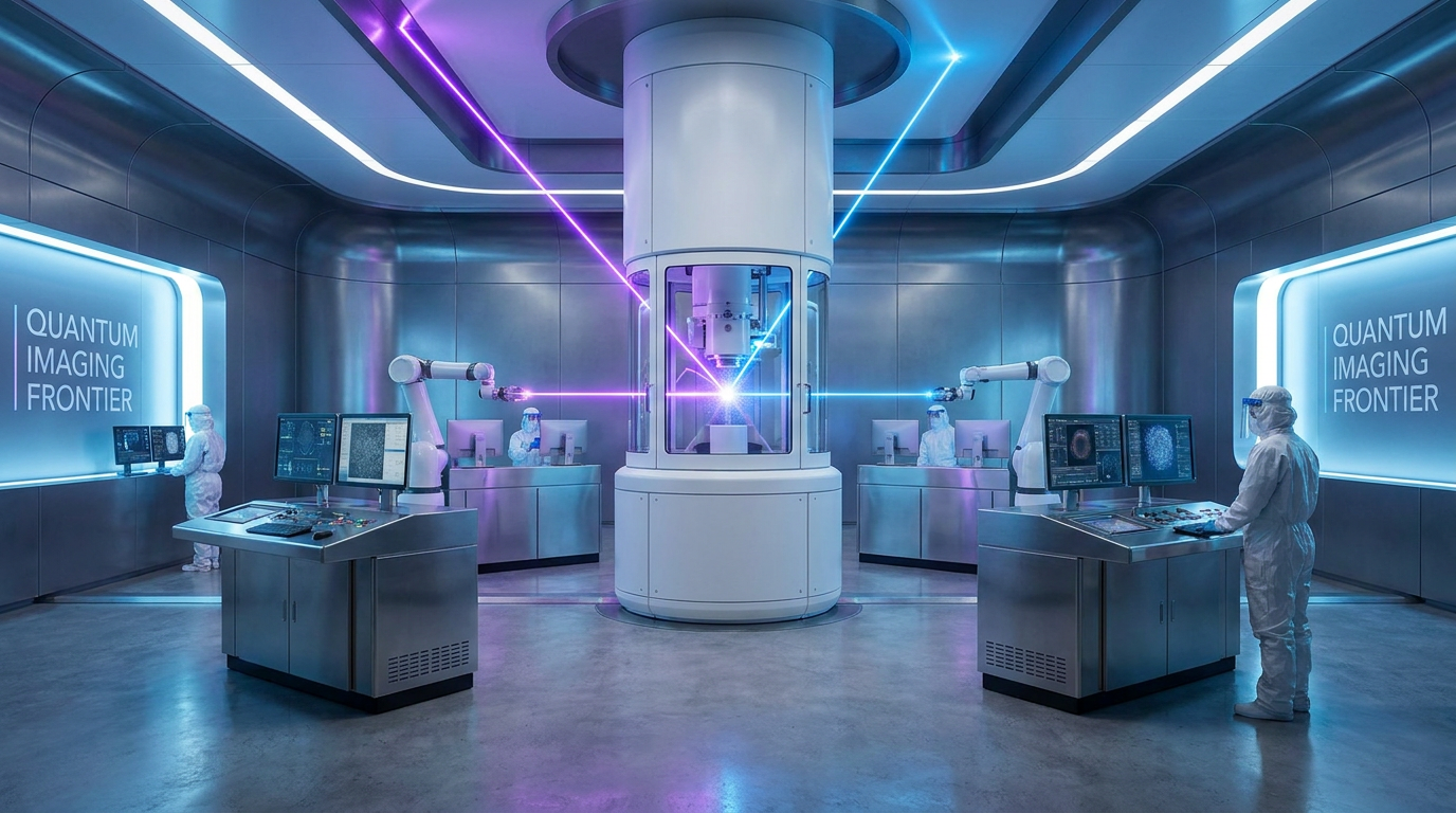
That fundamental limitation is now poised to fall. In a coordinated release of papers across major scientific journals, researchers from the University of California, Berkeley, and the Chan Zuckerberg Biohub have announced the successful demonstration of a "laser phase plate." The device, integrated into a state-of-the-art cryo-electron microscope, uses an intensely focused laser to dramatically boost the contrast of the images it produces. After 15 years of being dismissed by many as theoretically elegant but practically impossible to build, the technology has produced its first high-resolution images of previously elusive biological structures.[1][2][3]
To understand the breakthrough, one must look at the current gold standard for structural biology: cryo-electron microscopy (cryo-EM). The technique, which won the Nobel Prize in Chemistry in 2017, involves flash-freezing protein samples in a thin layer of vitreous ice and firing a beam of electrons through them. Because electrons have a much shorter wavelength than visible light, they can resolve individual atoms. A detector captures the electrons that pass through, and software stitches thousands of these 2D "shadows" into a 3D model.[4][6]
The problem lies in the physics of contrast. Biological molecules are composed of light elements like carbon, nitrogen, and oxygen, which do not scatter electrons very strongly. When the electron beam passes through a small protein, the resulting image is incredibly faint—akin to looking for a clear glass bead submerged in a glass of water. To generate enough contrast to see anything at all, microscopists have traditionally had to throw the image slightly out of focus, a compromise that inherently destroys the highest-resolution details.[3][7]
The theoretical solution has been known for nearly a century: phase contrast. By shifting the phase of the unscattered electrons by exactly 90 degrees, they can be made to interfere with the scattered electrons, converting invisible phase differences into stark, high-contrast amplitude differences. While physical phase plates made of carbon have been tried in electron microscopes, they quickly accumulate static electric charge from the electron beam, degrading the image and rendering them useless for long data-collection runs.[2][6]

The Berkeley team, led by physicist Holger Müller, hypothesized that a laser could act as an invisible, indestructible phase plate. Because light consists of electromagnetic waves, an intense enough laser field can exert a "ponderomotive force" on passing electrons, slowing them down just enough to shift their phase without physically touching or scattering them. The engineering hurdle was the sheer amount of light required: the laser needed to be roughly 100 million times brighter than the surface of the sun, contained within the vacuum of an electron microscope.[2][3][4]
The evidence published in Science confirms that the team successfully built this optical cavity. Using a custom-modified Thermo Fisher Krios microscope dubbed "Theia," the researchers bounced a continuous-wave laser between two highly polished mirrors, building up a massive standing wave of light directly in the path of the electron beam. The results provide strong evidence that the mechanism works exactly as predicted by quantum mechanics, delivering a stable, tunable 90-degree phase shift with no electrostatic charging.[2][6]
The evidence published in Science confirms that the team successfully built this optical cavity.
To prove the system's biological utility, the Berkeley team imaged hemoglobin, the oxygen-carrying protein in red blood cells. At roughly 64 kilodaltons, hemoglobin sits at the absolute lower limit of what conventional cryo-EM can resolve. With the laser phase plate turned on, the contrast improved dramatically. In paired experiments, the resolution of the hemoglobin structure improved by up to 44 percent, allowing the software to build a much more accurate atomic model from the faint signals.[3][7]

Crucially, the evidence shows that the laser does not degrade high-resolution data. When the team imaged aldolase, a larger benchmark enzyme that is relatively easy to capture with standard cryo-EM, the laser phase plate still improved the resolution. This confirms that the technology is strictly additive, boosting the low-frequency contrast (the overall shape and visibility of the molecule) without blurring the high-frequency details (the exact positions of individual atoms).[2][7]
While the single-laser system proved the concept, a parallel effort by the CZ Biohub team has already pushed the engineering further. In a preprint posted to bioRxiv, the Biohub researchers detailed a second-generation "crossed laser phase plate" (xLPP). This design uses two orthogonal laser beams arranged in an X-shape. The dual-beam approach splits the required optical power between two cavities, lowering the stress on the mirrors and reducing the risk of catastrophic thermal failure.[4][5][7]
The xLPP design also solves a subtle optical artifact. A single laser beam creates a slight asymmetry in the phase shift, which can produce faint "ghost images" that overlap with the real protein structure. The crossed lasers cancel out this asymmetry. Using the xLPP, the Biohub team achieved a staggering 1.79 Ångström resolution on apoferritin, a standard calibration protein. This near-atomic resolution proves that the dual-laser system can match the stringent quality standards required by modern structural biology.[5][7]

The most profound impact of the laser phase plate, however, will likely not be in imaging isolated proteins, but in a rapidly emerging field called cryo-electron tomography (cryo-ET). Instead of purifying proteins and studying them in a vacuum, cryo-ET aims to image intact, flash-frozen cells. By tilting the cellular sample and taking pictures from multiple angles, scientists can reconstruct a 3D volume of the cell, revealing how different proteins interact with each other in their native, crowded environment.[1][4]
Because intact cells are much thicker than isolated proteins, the electron beam scatters heavily, resulting in an incredibly noisy image where individual proteins are nearly impossible to spot. The Biohub team's preprint provides early evidence that the laser phase plate can cut through this noise. When imaging thick E. coli cells (roughly 350 nanometers deep), the xLPP provided a massive contrast boost, making internal cellular structures pop out clearly against the background.[5][7]

Despite the clear success of the prototypes, there is transparent uncertainty regarding how quickly this technology will reach the broader scientific community. The current systems are highly bespoke, requiring custom-machined microscope columns and constant tuning by expert optical physicists. As Holger Müller noted regarding the Berkeley prototype, "Ours is not street legal." The optical cavities require extreme precision; if the mirrors drift by even a fraction of a nanometer, the phase shift is lost.[6][7]
The next major hurdle is commercialization. Researchers are currently collaborating with microscope manufacturers like Thermo Fisher Scientific to determine if the laser phase plate can be miniaturized and stabilized into a plug-and-play module. Until it can be mass-produced and operated by standard facility technicians rather than dedicated physicists, its impact will be confined to a few elite imaging centers.[6]
If those engineering challenges are met, the laser phase plate represents a generational leap for biology. By illuminating the 90 percent of the proteome that has remained hidden, and by allowing scientists to watch those proteins operate inside the living machinery of the cell, the technology provides a new foundational map for human health. As Biohub's imaging director Bridget Carragher summarized, "It's like seeing first light through a telescope. The science it enables—that comes next."[4][7]
How we got here
1953
The Nobel Prize in Physics is awarded for the invention of the phase-contrast optical microscope.
2017
The Nobel Prize in Chemistry is awarded for the development of cryo-electron microscopy (cryo-EM).
2019
UC Berkeley researchers publish early proof-of-concept data showing a laser can shift the phase of an electron beam.
June 2026
Researchers publish coordinated papers demonstrating a working laser phase plate integrated into a state-of-the-art cryo-EM system.
Viewpoints in depth
Structural Biologists
Focused on resolving the atomic structures of small, elusive proteins to accelerate targeted drug discovery.
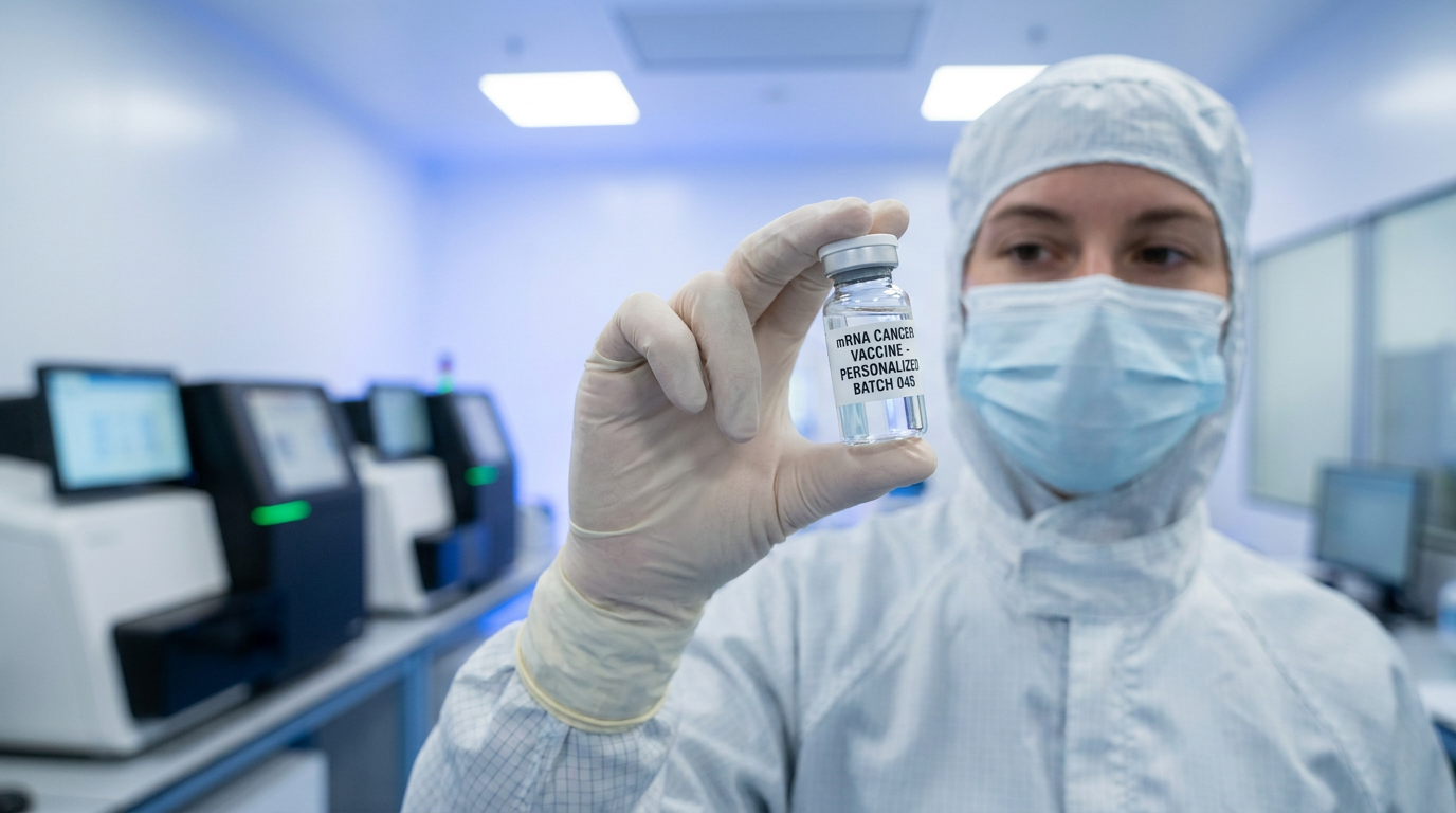
For structural biologists, the laser phase plate solves the field's most frustrating bottleneck: the inability to see the vast majority of the human proteome. Because standard cryo-EM relies on large proteins scattering enough electrons to create a shadow, researchers have historically been limited to studying massive molecular complexes. The ability to clearly image a 64-kilodalton protein like hemoglobin—and potentially much smaller targets—means that thousands of previously 'undruggable' proteins can now be mapped atom by atom. This camp views the technology primarily as an engine for pharmaceutical discovery, allowing them to design highly specific molecules that bind to newly revealed cellular targets.
Cellular Imagers
Prioritize the development of cryo-electron tomography to map how proteins interact dynamically inside intact cells.
Cellular imagers see the laser phase plate as the key to unlocking cryo-electron tomography (cryo-ET). While structural biologists want to study purified proteins in isolation, cellular imagers want to see the entire factory floor. Intact cells are thick and crowded, creating an incredibly noisy background that obscures individual proteins. The massive contrast boost provided by the laser phase plate cuts through this noise, allowing researchers to spot specific molecular machines operating in their native environment. For this group, the ultimate prize is building a complete, high-resolution 3D atlas of a living cell, showing exactly how proteins interact, assemble, and malfunction during disease.
Microscopy Engineers
Focused on the immense technical challenges of stabilizing optical cavities and scaling bespoke prototypes into commercial instruments.
For the physicists and engineers who built the system, the triumph is proving that a 15-year theoretical quest was actually possible. Building an optical cavity that sustains a laser 100 million times brighter than the sun inside the vacuum of an electron microscope requires borderline-impossible tolerances. Their focus is now on the harsh realities of commercialization. The current prototypes require constant, expert tuning to prevent the mirrors from drifting by even a fraction of a nanometer. This camp is working closely with manufacturers to determine if the delicate 'crossed laser' setup can be hardened into a stable, push-button module that standard laboratory technicians can operate without a PhD in optics.
What we don't know
- How long it will take to miniaturize and commercialize the laser phase plate for widespread laboratory use.
- Whether the dual-laser xLPP design will become the industry standard over the single-laser approach.
- The exact upper limit of resolution the laser phase plate can achieve on the smallest human proteins.
Key terms
- Cryo-electron microscopy (cryo-EM)
- An imaging technique that fires electrons at flash-frozen biological samples to determine the 3D atomic structure of molecules.
- Phase contrast
- An optical trick that converts invisible shifts in a wave's phase (timing) into visible differences in brightness and darkness, greatly enhancing image clarity.
- Ponderomotive force
- A nonlinear force exerted by an intense electromagnetic field (like a laser) that pushes charged particles, used here to shift the phase of the electron beam.
- Cryo-electron tomography (cryo-ET)
- An advanced imaging method that takes multiple pictures of an intact cell from different angles to build a 3D map of its internal environment.
- Kilodalton (kDa)
- A unit of mass used to measure the size of proteins and other large biological molecules.
- Ångström (Å)
- A unit of length equal to one ten-billionth of a meter, commonly used to measure atomic-level resolution in structural biology.
Frequently asked
What is cryo-electron microscopy (cryo-EM)?
Cryo-EM is a Nobel Prize-winning technique that flash-freezes biological samples and uses a beam of electrons to capture high-resolution 2D images, which are then assembled into 3D models of molecules.
Why couldn't standard cryo-EM see small proteins?
Small proteins are made of light elements that don't scatter electrons strongly enough to create a high-contrast image, making them appear nearly invisible against the background noise.
How does the laser phase plate fix this?
By shining an intensely bright laser across the electron beam, the light's electromagnetic field slows the electrons slightly, shifting their phase. This creates a high-contrast image without physically blocking or scattering the beam.
When will this be available in standard labs?
The technology is currently in the prototype phase and requires custom-built microscopes. Researchers are working with manufacturers to develop commercial, plug-and-play versions, but a timeline is not yet set.
Sources
Source coverage
7 outlets
3 viewpoints surfaced


[1]NatureStructural Biologists
An innovative technology boosts image quality for protein structures
Read on Nature →[2]ScienceStructural Biologists
Laser-boosted microscopes could reveal new drug targets, sharpen views inside cells
Read on Science →[3]UC Berkeley NewsMicroscopy Engineers
Physicists introduce phase contrast to electron microscopy, delivering sharper images of our body's tiniest proteins
Read on UC Berkeley News →[4]CZ BiohubCellular Imagers
Making the invisible visible: A landmark achievement in microscopy
Read on CZ Biohub →[5]bioRxivCellular Imagers
A Crossed Laser Phase Plate for Cryogenic Electron Microscopy
Read on bioRxiv →[6]Phys.orgMicroscopy Engineers
Physicists introduce phase contrast to electron microscopy
Read on Phys.org →[7]LabcriticsMicroscopy Engineers
Laser Phase Plate: The 'Impossible' Cryo-EM Upgrade is Here
Read on Labcritics →
Every angle. Every day.
Get science stories with full source coverage and perspective breakdowns delivered to your inbox.