A 75-Kilowatt Laser Just Solved Cryo-Electron Microscopy's Biggest Blind Spot
By trapping a laser 100 million times brighter than the sun inside an electron microscope, physicists have dramatically boosted image contrast, allowing scientists to finally see the smallest proteins in the human body.
By Factlen Editorial Team
- Structural Biologists
- Value the technology for its potential to reveal the remaining 90% of the human proteome and accelerate drug discovery.
- Optical Physicists
- Focus on the engineering milestone of successfully manipulating an electron beam with a high-intensity continuous laser.
- Microscopy Core Directors
- Evaluate the practical deployment, stability, and daily operational impact of integrating the laser into existing lab facilities.
What's not represented
- · Commercial microscope manufacturers
- · AI structure-solving software developers
Why this matters
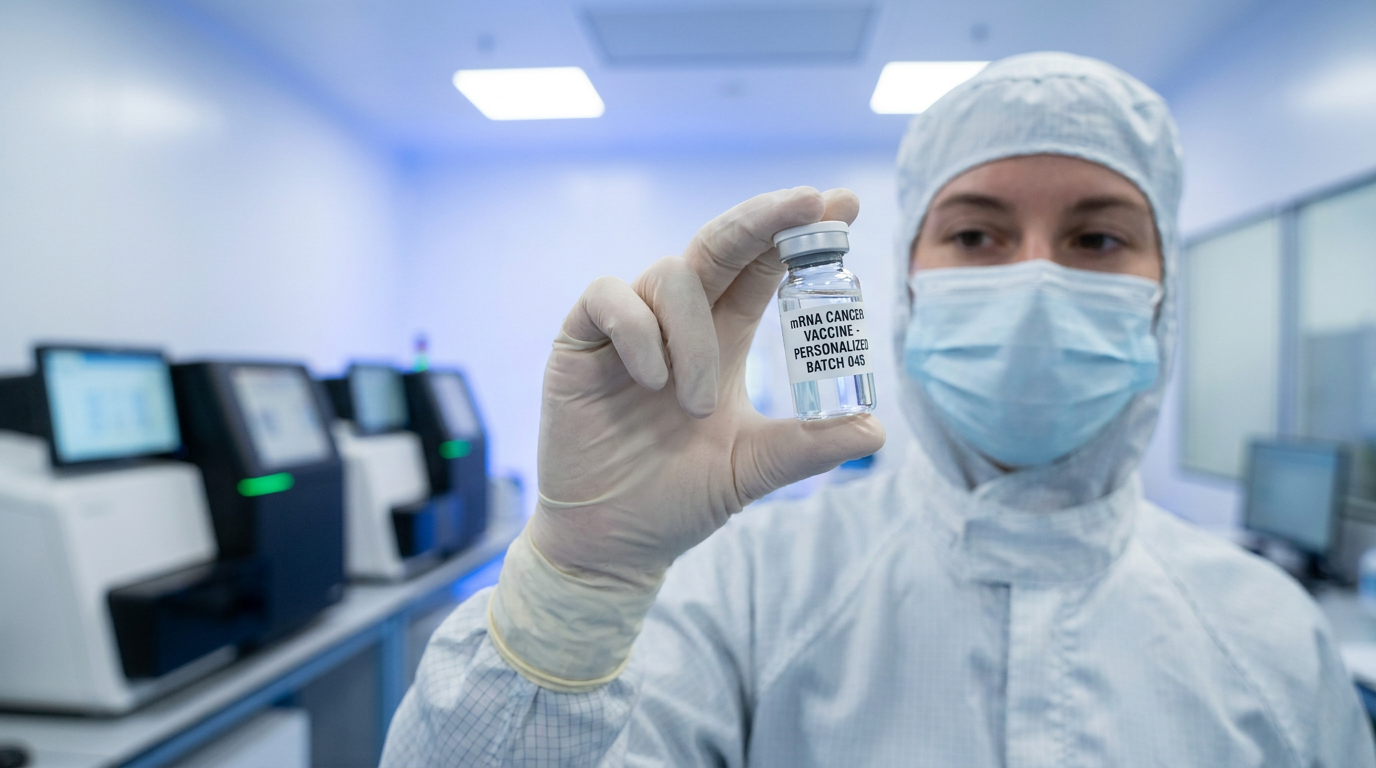
More than 90 percent of the proteins inside human cells are currently too small to be seen by electron microscopes. By using a laser to dramatically boost image contrast, this breakthrough allows scientists to finally visualize the hidden molecular machinery that drives both fundamental biology and complex diseases, accelerating the discovery of new life-saving drugs.
Key points
- Cryo-electron microscopy struggles to image small proteins because biological samples are essentially transparent to electron beams.
- Physicists successfully trapped a 75-kilowatt continuous laser inside a microscope to shift the phase of the electron beam.
- The laser phase plate dramatically boosts image contrast without relying on physical materials that degrade or charge.
- The technology successfully imaged hemoglobin and apoferritin at high resolutions, proving its efficacy on small molecules.
- Researchers believe the device will revolutionize cryo-electron tomography by allowing scientists to map proteins inside intact cells.
Cryo-electron microscopy is one of the most powerful tools in modern science, earning a Nobel Prize for its ability to reveal the atomic architecture of the biological machines that keep us alive. By firing a beam of electrons at flash-frozen samples, researchers can determine the three-dimensional shapes of viruses, enzymes, and cellular receptors. Yet, despite its revolutionary impact on drug discovery and structural biology, the technique has harbored a massive blind spot. Because biological molecules are composed of light elements like carbon and oxygen, they are essentially transparent to electron beams. To generate a visible image, conventional cryo-electron microscopy relies on a delicate optical compromise that inherently sacrifices contrast. As a result, the technology has historically been restricted to imaging large, massive protein complexes. Researchers estimate that more than 90 percent of the proteins found inside human cells are simply too small to generate enough contrast to be captured clearly, leaving the vast majority of our cellular machinery hidden in the dark.[1][4]
On June 11, 2026, a coalition of researchers from the University of California, Berkeley, and the Chan Zuckerberg Biohub announced they had successfully engineered a solution to this limitation. Across a coordinated series of publications in leading journals, the teams demonstrated a working "laser phase plate"—a novel device that uses an intensely focused beam of light to dramatically boost the contrast of electron microscopes. By integrating this optical cavity into a state-of-the-art microscope, the researchers successfully imaged molecules that were previously considered at the absolute lower limit of the technology's capabilities. The breakthrough promises to open an entirely new window into human biology, allowing scientists to visualize the faint, blurry proteins that drive both fundamental cellular processes and complex diseases.[1][3][4]
The primary claim across the newly published research is that the laser phase plate provides a stable, tunable contrast enhancement that allows cryo-electron microscopy to resolve small molecules without compromising high-resolution data. To understand the weight of this evidence, one must look back at an 80-year-old physics problem. In 1942, Dutch physicist Fritz Zernike discovered that inserting a physical plate into a light microscope could shift the phase of scattered light waves. This optical trick converted invisible shifts in a wave's timing into highly visible differences in brightness, making transparent biological cells vivid and earning Zernike the 1953 Nobel Prize in Physics. For decades, structural biologists attempted to replicate Zernike's phase-contrast trick in electron microscopes using physical material plates.[6][7]

The evidence consistently showed that these physical phase plates failed in practice. When placed in the path of an electron beam, thin films of carbon or other materials would rapidly accumulate electrostatic charge. This charging effect acted like a distorted lens, scrambling the electron waves and ruining the high-resolution information required to build atomic models. More than 15 years ago, UC Berkeley physicists Holger Müller and Robert Glaeser proposed a theoretical alternative: instead of using a solid material that could charge or degrade, why not use a concentrated beam of light to shift the electrons' phase? Because photons do not carry an electric charge, a laser could theoretically provide the necessary phase shift through the ponderomotive potential—a phenomenon where electrons are repelled by a high-intensity electromagnetic field—without introducing any physical contamination.[2][4][6]
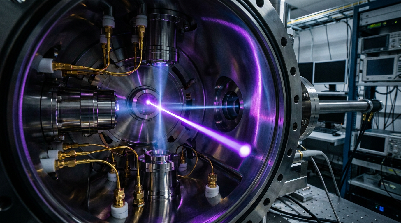
While theoretically elegant, the engineering required to test this claim was widely dismissed by the field as practically impossible. To shift the phase of a 300-kilovolt electron beam by the necessary 90 degrees, the laser would need to be unimaginably powerful. The UC Berkeley team spent more than a decade building a device capable of trapping a continuous-wave laser inside a spherical, mirrored cavity. By bouncing the light back and forth between highly polished mirrors more than 10,000 times, the cavity amplifies the beam to 75 kilowatts of power, focused down to a spot just a few micrometers wide. This creates a steady-state light field roughly 100 million times brighter than the sun.[3][4]
The sheer physical constraints of the project added to the difficulty. The entire optical cavity housing this immensely powerful laser had to be miniaturized to less than four inches wide so it could be tucked inside the vacuum column of a 14-foot-tall Thermo Fisher Scientific Krios microscope. The evidence for the single-laser phase plate's efficacy is robustly documented in the team's Science publication. The researchers presented reconstructed images of several biological samples, including aldolase—a muscle protein that is relatively easy to image—and hemoglobin, the oxygen-carrying protein in blood that represents the lower size limit for current machines.[2][3][4]

The sheer physical constraints of the project added to the difficulty.
The data demonstrated a clear, quantifiable improvement. The laser phase plate improved the resolution of the protein structures in both cases, but the evidence showed that the smaller the sample, the greater the contrast enhancement provided by the laser. This directly validated the theoretical models proposed by Müller and Glaeser years earlier. By converting the phase modulation imprinted by the specimen into detectable amplitude modulation, the laser phase plate allowed the structure-solving software to generate highly accurate atomic models from images that would have otherwise been too washed out to process.[2][3][7]
Meanwhile, a parallel engineering effort at CZ Biohub developed a second-generation design known as the crossed laser phase plate, or xLPP, which is detailed in a newly released bioRxiv preprint. Rather than relying on a single optical cavity, the xLPP utilizes two intersecting laser beams arranged in an orthogonal, X-shaped configuration. The researchers claim this dual-beam approach offers two concrete advantages over the single-laser design. First, it splits the required power between two sets of mirrors, meaning each cavity runs at roughly half the intensity. This reduces the thermal stress on the components and lowers the risk of catastrophic optical failure.[5][6]
Second, the crossed configuration effectively suppresses "ghost images." In phase-contrast microscopy, shifting the beam can sometimes create faint, duplicate copies of high-contrast objects in the background, which can swamp the delicate biological signals researchers are trying to isolate. The evidence for the xLPP's performance is strong for purified proteins. In their preprint, the Biohub team demonstrated that single-particle analysis of apoferritin—a standard iron-storage protein used as a benchmark in the field—reached a resolution of 1.79 angstroms. This proves that the dual-laser system is capable of acquiring the high-resolution data required for modern drug design and structural mapping.[5][6]

However, the most highly anticipated claim surrounding the laser phase plate is its potential to revolutionize a newer technique known as cryo-electron tomography, or cryo-ET. While standard cryo-electron microscopy studies purified proteins isolated in a solution, cryo-ET captures multiple angular views of an intact, flash-frozen cell to build a three-dimensional map of its interior. This allows scientists to analyze proteins in their natural, crowded environments, observing how molecular machines interact with their neighbors to carry out the processes of life. Because intact cells are much thicker than purified protein samples, the contrast problem is exponentially worse, making the laser phase plate a critical necessity for the field's advancement.[1][4][7]
The evidence supporting the laser phase plate's utility in cryo-ET remains in the early, proof-of-concept stages. The bioRxiv preprint demonstrates that when imaging thick Escherichia coli cells—measuring roughly 350 nanometers deep—the crossed laser phase plate successfully enhanced the overall contrast. The researchers noted a measurable increase in the low-frequency signal, which is essential for template-matching algorithms to identify specific proteins within the dense cellular soup. While this confirms the physics work in thicker samples, the comprehensive mapping of a human cell's interior remains a future goal rather than a completed milestone.[5][6]
Despite the overwhelming success of the physical instrument, researchers are transparent about the uncertainties and the work that lies ahead. As Biohub's technical director of imaging noted, the current achievement is akin to seeing "first light through a telescope." The physical hardware works exactly as the theoretical physics predicted, but the technology is not yet a plug-and-play solution ready for deployment in every university core facility. Maintaining the precise alignment of a 75-kilowatt laser inside a vacuum chamber requires specialized expertise, and the optical cavities will need further refinement to ensure long-term stability and commercial scalability.[4][6]

Furthermore, the influx of high-contrast data will require parallel advancements in computational biology. As the microscopes begin capturing clearer images of smaller, highly dynamic proteins, the artificial intelligence algorithms used to reconstruct these 3D models will need to adapt to the new wealth of information. The next phase of evidence gathering will inevitably shift from physics to biology. With the contrast barrier finally broken, structural biologists are preparing to turn these upgraded microscopes toward the 90 percent of the human proteome that has remained invisible, searching for the molecular roots of neurodegeneration, cancer, and infectious diseases.[1][4]
Ultimately, the realization of the laser phase plate marks the end of an 80-year quest to bring Zernike's vision to the electron scale. By replacing physical materials with the pure ponderomotive potential of light, physicists have decoupled image contrast from resolution loss. As these instruments transition from custom-built prototypes to standardized laboratory tools, the scientific community anticipates a renaissance in structural biology. The ability to routinely visualize the smallest cogs of the cellular machine will not only rewrite biology textbooks but also provide pharmaceutical researchers with the precise atomic blueprints needed to design the next generation of targeted therapeutics.[2][4][7]
How we got here
1942
Fritz Zernike invents phase-contrast for light microscopes, an achievement that later wins the 1953 Nobel Prize.
2010
UC Berkeley physicists Holger Müller and Robert Glaeser publish the theoretical framework for a laser phase plate in electron microscopes.
2021
CZ Biohub provides a major grant to accelerate the physical construction of the laser phase plate prototype.
June 2026
Researchers publish multiple papers demonstrating working single-laser and crossed-laser phase plates, achieving unprecedented contrast for small proteins.
Viewpoints in depth
Structural Biologists
Focused on the biological applications and the ability to map the human proteome.
For structural biologists, the laser phase plate is the key to unlocking the 'dark matter' of the cell. Because conventional cryo-EM struggles with molecules under a certain weight threshold, researchers have historically been forced to study only the largest, most stable protein complexes. By boosting contrast at the hardware level, biologists argue they can now visualize the remaining 90 percent of human proteins, accelerating drug discovery and mapping cellular machinery in its native state.
Optical Physicists
Focused on the engineering triumph of manipulating electron beams with light.
From a physics perspective, the achievement lies in overcoming a barrier deemed practically impossible for 15 years. Physicists emphasize the sheer difficulty of trapping a 75-kilowatt continuous laser in a four-inch cavity without destroying the surrounding equipment. They view the successful deployment of both the single and crossed laser phase plates as a landmark validation of ponderomotive potential theories, proving that light can serve as a flawless, non-degrading optical element for electron beams.
What we don't know
- How quickly the laser phase plate can be commercialized and deployed to standard university core facilities.
- Whether the current artificial intelligence algorithms used for structure-solving can efficiently handle the new influx of high-contrast cellular data.
- The exact timeline for achieving a complete, high-resolution 3D map of an intact human cell using this technology.
Key terms
- Cryo-electron microscopy (cryo-EM)
- A technique used to image flash-frozen molecules at atomic resolution using a beam of electrons.
- Phase contrast
- An optical trick that converts invisible shifts in a wave's timing (phase) into visible differences in brightness (amplitude).
- Proteome
- The entire set of proteins expressed by a genome, cell, tissue, or organism at a certain time.
- Angstrom (Å)
- A unit of length equal to one ten-billionth of a meter, commonly used to measure atomic-level resolution in structural biology.
- Cryo-electron tomography (cryo-ET)
- An advanced imaging technique that captures multiple angled views of a frozen cell to build a 3D model of its internal structure.
Frequently asked
What is cryo-electron microscopy?
A Nobel-winning scientific technique that fires electrons at flash-frozen biological samples to determine their three-dimensional atomic structures.
Why was a laser added to the microscope?
Biological samples are mostly transparent to electrons. The laser shifts the phase of the electron beam, creating high-contrast images of tiny proteins that were previously invisible.
Can this technology see inside living cells?
Not living cells, as samples must be flash-frozen. However, it enables cryo-electron tomography (cryo-ET), which maps proteins in their natural, intact cellular environments rather than in isolated solutions.
Sources
Source coverage
7 outlets
3 viewpoints surfaced


[1]NatureStructural Biologists
An innovative technology boosts image quality for protein structures
Read on Nature →[2]ScienceOptical Physicists
Laser phase contrast for cryo-electron microscopy
Read on Science →[3]UC Berkeley NewsMicroscopy Core Directors
A breakthrough in electron microscopy delivers sharper images of our body's tiniest proteins
Read on UC Berkeley News →[4]CZ BiohubStructural Biologists
Microscope Breakthrough Will Open Unprecedented View into Our Cells
Read on CZ Biohub →[5]bioRxivOptical Physicists
A Crossed Laser Phase Plate for CryoEM
Read on bioRxiv →[6]LabCriticsMicroscopy Core Directors
The Laser Phase Plate is Finally Here
Read on LabCritics →[7]Phys.orgOptical Physicists
Physicists introduce phase contrast to electron microscopy, delivering sharper images of our body's tiniest proteins
Read on Phys.org →
Every angle. Every day.
Get science stories with full source coverage and perspective breakdowns delivered to your inbox.