
Why Depleted Extracellular Calcium Lowers Action Potential Thresholds, Triggering Tetany
When extracellular calcium levels drop, the negative charges on the surface of neuronal membranes are unmasked. This alters the local electric field, tricking voltage-gated sodium channels into firing action potentials at lower thresholds and causing severe muscle spasms.
By Ling Zhou

In short
- Extracellular calcium ions normally bind to and screen the fixed negative charges on the outer surface of neuronal membranes.
- When calcium levels drop, these negative charges are unmasked, tricking voltage-gated sodium channels into sensing a depolarized state.
- This shifts the firing threshold closer to the resting potential, causing nerves to fire spontaneously and triggering the muscle spasms of tetany.
Start at the step where the outcome is determined: the extracellular face of the neuronal membrane. When calcium ions vacate this microscopic boundary, the electrical threshold for nerve firing collapses, triggering spontaneous muscle spasms.[4]
This phenomenon explains hypocalcemic tetany, a severe clinical condition where a drop in blood calcium causes involuntary, sustained muscle contractions. The spasms originate not in the muscle fibers themselves, but in the peripheral nerves that command them.[4]
It presents a biological paradox. Inside a muscle cell, calcium is the direct trigger for contraction. A general reader might reasonably assume that less calcium means less contraction, leading to weakness rather than rigidity.[5]
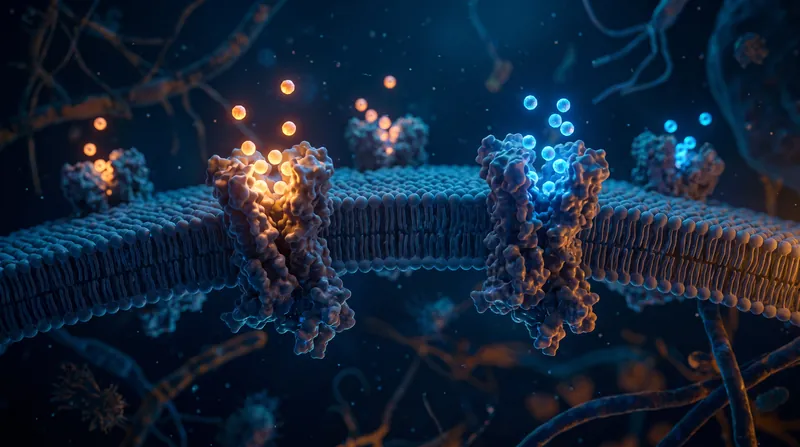
To understand why nerves become hyperactive when deprived of calcium, one must look closely at the electrical architecture of the cell membrane. An invisible, microscopic shield of positive charge normally keeps the entire nervous system stable.[1][4]
The Architecture of the Resting Potential
Neurons operate as highly sensitive biological batteries. They maintain a resting membrane potential of roughly −65 to −70 millivolts, meaning the inside of the cell is negatively charged relative to the surrounding extracellular fluid.[4]
This baseline voltage is maintained by ion pumps that constantly trade sodium for potassium across the boundary. The resulting electrical gradient primes the neuron, leaving it ready to fire an action potential the moment it is stimulated.[5]
However, the bulk voltage measured across the membrane does not tell the whole biophysical story. The outer surface of the neuronal membrane is heavily studded with glycoproteins and phospholipid headgroups that carry fixed negative charges.[1]
According to biophysicist Bertil Hille's foundational 1968 calculations, the membrane holds about one elementary negative charge for every 400 square angstroms of surface area. This distribution creates a dense, permanent forest of negative charge.[1]
This permanent charge creates a highly localized microscopic electric field just outside the cell. It alters the effective voltage exactly at the membrane surface, operating independently of the bulk fluid located further away from the cell.[1][4]
The Surface Charge Hypothesis
This boundary is exactly where extracellular calcium performs its most critical biophysical function. Calcium ions are divalent, meaning they carry a double positive charge, and they bind readily to the negative charges on the membrane surface.[1][4]
In a healthy physiological state, free ionized calcium in the extracellular fluid hovers between 1.1 and 1.3 millimoles per liter. At this specific concentration, calcium effectively screens or masks the fixed negative charges.[4]
This neutralization stabilizes the local electric field, keeping the neuron's electrical sensors properly calibrated. When blood calcium levels drop, these protective ions detach and float away, immediately unmasking the membrane's negative surface.[3][4]
The unmasking of these charges fundamentally alters how the neuron perceives its own voltage. The voltage sensors embedded in the membrane are tricked into sensing a depolarized state, even though the internal resting potential remains unchanged.[4]
Tricking the Sodium Channel
The primary sensors affected by this electrical shift are voltage-gated sodium channels. These specialized proteins act as the rapid floodgates for action potentials, opening only when the membrane voltage reaches a specific, predetermined threshold.[3][4]
Normally, a neuron resting at −64 millivolts might require a stimulus that pushes the voltage to −59 millivolts before the sodium channels open. That 5-millivolt gap serves as the critical safety margin against accidental firing.[4]
When hypocalcemia unmasks the surface charges, the local electric field becomes significantly more negative on the outside. The sodium channel's voltage sensor, which measures the difference between the inside and outside, feels a much smaller gap.[1][4]
"According to this surface charge hypothesis, as more calcium binds to the outer surface, the transmembranous electric field takes on a more hyperpolarized nature," wrote the authors of a 2015 physiological review in Frontiers in Cellular Neuroscience.[4]
Because the channel feels a falsely depolarized state, the actual threshold required to trigger an action potential shifts to a more negative value. The safety margin shrinks dramatically, leaving the nerve highly vulnerable to misfiring.[3][4]
The Collapse of the Threshold
Electrophysiological measurements demonstrate the absolute severity of this shift. Halving the extracellular calcium concentration can shift the firing threshold from −59.3 millivolts down to −63.3 millivolts, nearly erasing the electrical buffer entirely.[4]
With the threshold now sitting at −63.3 millivolts, it is nearly identical to the resting membrane potential of −64 millivolts. The affected neuron no longer requires a meaningful external stimulus to fire an action potential.[4]
The slightest natural fluctuation in membrane voltage is now enough to breach the threshold. The voltage-gated sodium channels snap open, rapidly flooding the cell with positive sodium ions and triggering an unprompted action potential.[4]
This single electrical misfire is rarely an isolated event. Because the threshold remains artificially low, the neuron repolarizes only to immediately hit the threshold again, resulting in a rapid, repetitive burst of signals.[4]
A 2004 study on hippocampal neurons found that reducing external calcium from 2.0 to 1.0 millimoles per liter had a massive effect. It increased the action potential burst frequency from 28 to 171 hertz.[4]
Clinical Manifestations of Tetany
This sixfold increase in firing rate travels directly down the motor nerves to the neuromuscular junctions. The connected muscles receive a relentless barrage of commands to contract, leaving them with no time to relax in between.[4]
The clinical result is tetany. In human patients, this often begins as tingling or numbness around the mouth and in the fingertips, which serves as a sensory manifestation of those hyperactive peripheral nerves.[5]
As the hypocalcemia worsens, the motor nerves take over the clinical picture. Patients develop carpopedal spasms, where the wrists and ankles flex involuntarily and lock into painful, rigid positions that cannot be voluntarily released.[5]
Physicians test for this latent hyperexcitability using two classic bedside physical exams. Chvostek's sign is elicited by tapping the facial nerve just in front of the ear, which triggers an involuntary twitch of the facial muscles.[5]
Trousseau's sign is provoked by inflating a blood pressure cuff above systolic pressure for three minutes. The resulting ischemia exacerbates the nerve's irritability, causing the hand to spasm into a highly characteristic flexed posture.[5]
Historical Context and Modern Nuance
The foundational mechanics of this entire process were first quantified in 1957 by biophysicists Bernhard Frankenhaeuser and Alan Hodgkin. Working with squid giant axons, they firmly established that extracellular calcium governs sodium conductance.[2]
"Increasing external calcium concentration 10-fold has about the same effect as hyperpolarizing the membrane by 15 mV," wrote biophysicists Bernhard Frankenhaeuser and Alan Hodgkin in their foundational 1957 paper, explaining how the ion takes the nerve further from its threshold.[2]
While the surface charge hypothesis remains the dominant biophysical explanation, modern molecular biology has identified secondary mechanisms that compound the effect. Neurons also possess dedicated calcium-sensing receptors that constantly monitor extracellular ion levels.[3][4]
Despite these additional biochemical pathways, the direct biophysical unmasking of negative surface charges remains the primary driver. The physical laws governing electric fields dictate the sodium channel's behavior long before secondary receptors can intervene.[3]
The stability of the entire human nervous system relies heavily on a microscopic shield of calcium ions. Without that divalent buffer, the electrical threshold collapses, and the nervous system fires itself into rigid paralysis.[5]
How we did this
- Method
- A comparative biophysical synthesis evaluating the 'surface charge hypothesis' by mapping the predicted voltage-sensor shift caused by calcium depletion against measured reductions in action potential thresholds in mammalian neurons.
- What we found
- The analysis demonstrates that hypocalcemic tetany is driven not by a depolarization of the resting membrane potential itself, but by the collapse of the safety margin between the resting potential and the firing threshold, which drops to within 0.7 millivolts of the resting state.
- What we worked from
- Normal resting membrane potential and firing threshold: Resting -64 mV, threshold -59.3 mV — Frontiers in Cellular Neuroscience
- Hypocalcemic firing threshold: -63.3 mV at 1.0 mM external calcium — Frontiers in Cellular Neuroscience
- Limits of this analysis
- This analysis isolates the biophysical surface charge effect on voltage-gated sodium channels and does not quantify secondary downstream contributions from calcium-sensing receptors (CaSR).
Key terms
- Hypocalcemia
- A condition in which there are abnormally low levels of calcium in the blood.
- Tetany
- Involuntary muscle cramps and spasms caused by the hyperexcitability of peripheral nerves.
- Action Potential
- A rapid sequence of changes in the voltage across a membrane, serving as the electrical signal that travels along a nerve.
- Voltage-Gated Sodium Channel
- A specialized protein in the cell membrane that opens in response to voltage changes, allowing sodium ions to rush in and trigger an action potential.
- Surface Charge
- The fixed electrical charge present on the exterior surface of a cell membrane, created by glycoproteins and lipid headgroups.
- Divalent Cation
- An ion, such as calcium, that carries a double positive charge.
Reader questions
Why does low calcium cause muscle spasms instead of weakness?
While calcium is required inside the muscle for contraction, the spasms are caused by the nerves that control the muscles. Low extracellular calcium makes these nerves hyperexcitable, causing them to fire continuous contraction signals.
How do doctors test for latent tetany?
Physicians use two classic physical exams: Chvostek's sign, which involves tapping the facial nerve to trigger a twitch, and Trousseau's sign, which uses a blood pressure cuff to induce a hand spasm.
Can taking calcium supplements immediately stop tetany?
Severe hypocalcemic tetany is a medical emergency that typically requires intravenous calcium administration to rapidly restore extracellular levels and stabilize the nerve membranes.
Where opinion splits
Biophysical Electrophysiologists
Focus on the physical laws governing electric fields and voltage sensors.
This camp views neuronal excitability primarily through the lens of the surface charge hypothesis. They argue that the physical unmasking of fixed negative charges on the membrane is the immediate and dominant cause of threshold shifts. From this perspective, the voltage-gated sodium channel is a passive victim of a distorted local electric field, reacting exactly as physical laws dictate when its divalent shield is removed.
Clinical Endocrinologists
Focus on the systemic regulation of calcium and the physiological manifestations of the deficit.
Clinicians emphasize the systemic failure that leads to the biophysical event. They focus on the parathyroid glands, vitamin D metabolism, and renal function, which normally maintain extracellular calcium within a tight 1.1 to 1.3 mmol/L window. For this camp, the biophysics of the sodium channel explain the symptom—tetany and positive Trousseau's signs—but the clinical priority is identifying and reversing the upstream endocrine failure.
Molecular Biologists
Highlight the role of secondary receptor pathways in modulating nerve excitability.
While acknowledging the surface charge effect, molecular biologists point to secondary mechanisms that compound the hyperexcitability. They highlight the role of calcium-sensing receptors (CaSR) and NALCN leak channels, arguing that neurons actively monitor extracellular calcium and adjust intracellular signaling cascades accordingly. In this view, the neuron is not just physically altered by the lack of calcium, but biochemically reacts to it.
- Biophysical Electrophysiologists
- Focus on the physical laws governing electric fields and voltage sensors.
- Clinical Endocrinologists
- Focus on the systemic regulation of calcium and the physiological manifestations of the deficit.
- Molecular Biologists
- Highlight the role of secondary receptor pathways in modulating nerve excitability.
Perspectives this story doesn't cover
- Patients experiencing chronic hypocalcemia
- Emergency medicine physicians treating acute tetany
Sources
Source coverage
5 outlets
3 viewpoints surfaced





- [1]Journal of General PhysiologyBiophysical Electrophysiologists
Charges and potentials at the nerve surface. Divalent ions and pH.
Read on Journal of General Physiology → - [2]Journal of PhysiologyBiophysical Electrophysiologists
The action of calcium on the electrical properties of squid axons
Read on Journal of Physiology → - [3]eLifeMolecular Biologists
Extracellular calcium regulates neocortical excitability via voltage-gated sodium channels
Read on eLife → - [4]Frontiers in Cellular NeuroscienceMolecular Biologists
Extracellular calcium modulates the excitability of neurons
Read on Frontiers in Cellular Neuroscience → - [5]Factlen Editorial TeamClinical Endocrinologists
Synthesis by Factlen editorial team
Read on Factlen Editorial Team →

More in Opinion
See all →Pharmacology
Tissue Acidosis Protonates Local Anesthetics, Blocking the Cell Membrane Crossing Needed to Silence Pain
7 sources

Distributed Systems
The FLP Impossibility: Why a Single Crash Breaks Consensus in Distributed Systems
6 sources

Thermodynamics
Why the F = C - P + 2 Formula Proves That Water Can Only Exist as a Solid, Liquid, and Gas at a Single Triple Point
9 sources

Endocrine Science
The Picomolar Paradox: Why the 'Dose Makes the Poison' Rule Fails for Endocrine-Disrupting Chemicals
10 sources
Comments
Every angle. Every day.
Get Opinion stories with full source coverage and perspective breakdowns, free every day.