
Single-Cell Studies Reveal Massive Immune Cell Turnover and Genome Breakdown in the Aging Human Brain
New high-resolution mapping of the human brain reveals that its original immune cells are largely replaced by bone marrow-derived cells between the ages of 50 and 75. The findings fundamentally rewrite biological assumptions about brain aging and open new pathways for Alzheimer's treatments.
By Harper Lane

- Neuroimmunologists
- Argue that the influx of peripheral immune cells drives chronic neuroinflammation and exacerbates neurodegenerative diseases.
- Alzheimer's Researchers
- Suggest that marrow-derived macrophages may actually protect the brain by clearing toxic amyloid plaques more effectively than exhausted original microglia.
- Aging Biologists
- Focus on the fundamental breakdown of 3D genome architecture as the root cause of cellular confusion and aging in the brain.
Why this matters
Understanding how the brain ages is the critical first step to curing neurodegenerative diseases like Alzheimer's. The discovery that the brain's immune system is actively replaced by the bone marrow in later life opens up entirely new avenues for treatment, suggesting therapies could target the blood rather than the brain itself.
Key points
- The aging human brain undergoes a massive structural remodeling starting in midlife.
- Original brain immune cells are replaced by bone marrow-derived cells between ages 50 and 75.
- The 3D physical architecture of DNA breaks down across multiple brain cell types.
- The blood-brain barrier weakens with age, allowing peripheral immune cells to enter.
- Researchers debate whether these new immune cells drive inflammation or protect against Alzheimer's.
For decades, neuroscience textbooks have maintained a relatively static view of the adult brain's immune system. The prevailing consensus held that microglia—the brain's primary immune cells—are seeded during embryonic development and remain isolated behind the blood-brain barrier for a person's entire life.[1][6]
However, a wave of new high-resolution single-cell research has fundamentally rewritten this biological dogma. Rather than a slow, uniform decline, the aging human brain undergoes a massive, active structural remodeling starting in midlife.[2][6]
This evidence pack synthesizes findings from two landmark 2026 studies published in Science and Cell, which utilized advanced single-cell transcriptomics and somatic mutation tracking to map the aging brain. The data reveals that the brain's immune population is largely replaced, its physical DNA architecture erodes, and its protective barrier weakens.[1][3]
The most striking claim across these recent datasets is the wholesale turnover of microglia. Researchers mapping the human hippocampus observed a dramatic shift occurring primarily between the ages of 50 and 75, fundamentally altering the brain's cellular landscape.[1][2]
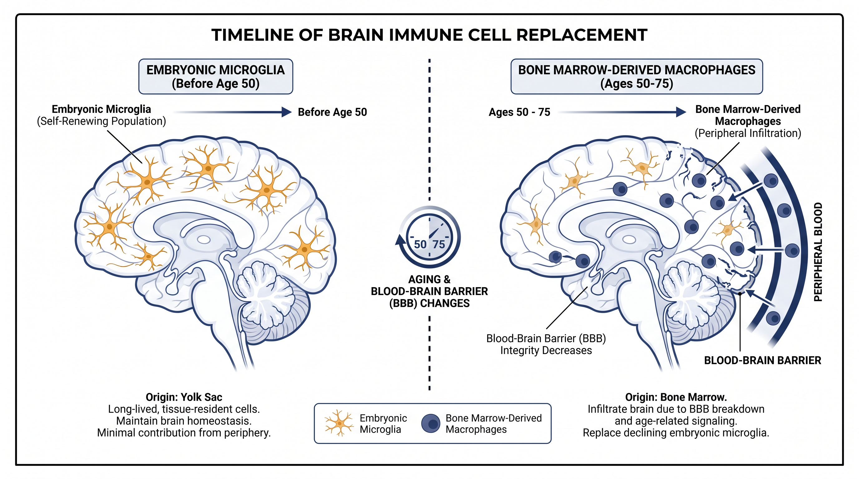
During this 25-year window, the microglia that originated during embryonic development decline sharply in number. In their place, a new population of cells emerges, possessing molecular signatures nearly identical to immune cells circulating in the bloodstream.[2]
The evidence for this infiltration is robustly supported by a separate study that tracked somatic mutations—natural DNA changes that accumulate over time and act as unique barcodes for cell lineages. By analyzing brain tissue from 20 older adults, researchers definitively traced these new brain macrophages back to the bone marrow.[3][4]
In all 20 individuals examined, marrow-derived cells had successfully crossed into the brain, taking up residence and adopting a microglia-like state. In some subjects, these infiltrating cells comprised a substantial majority of the total microglial pool.[4]
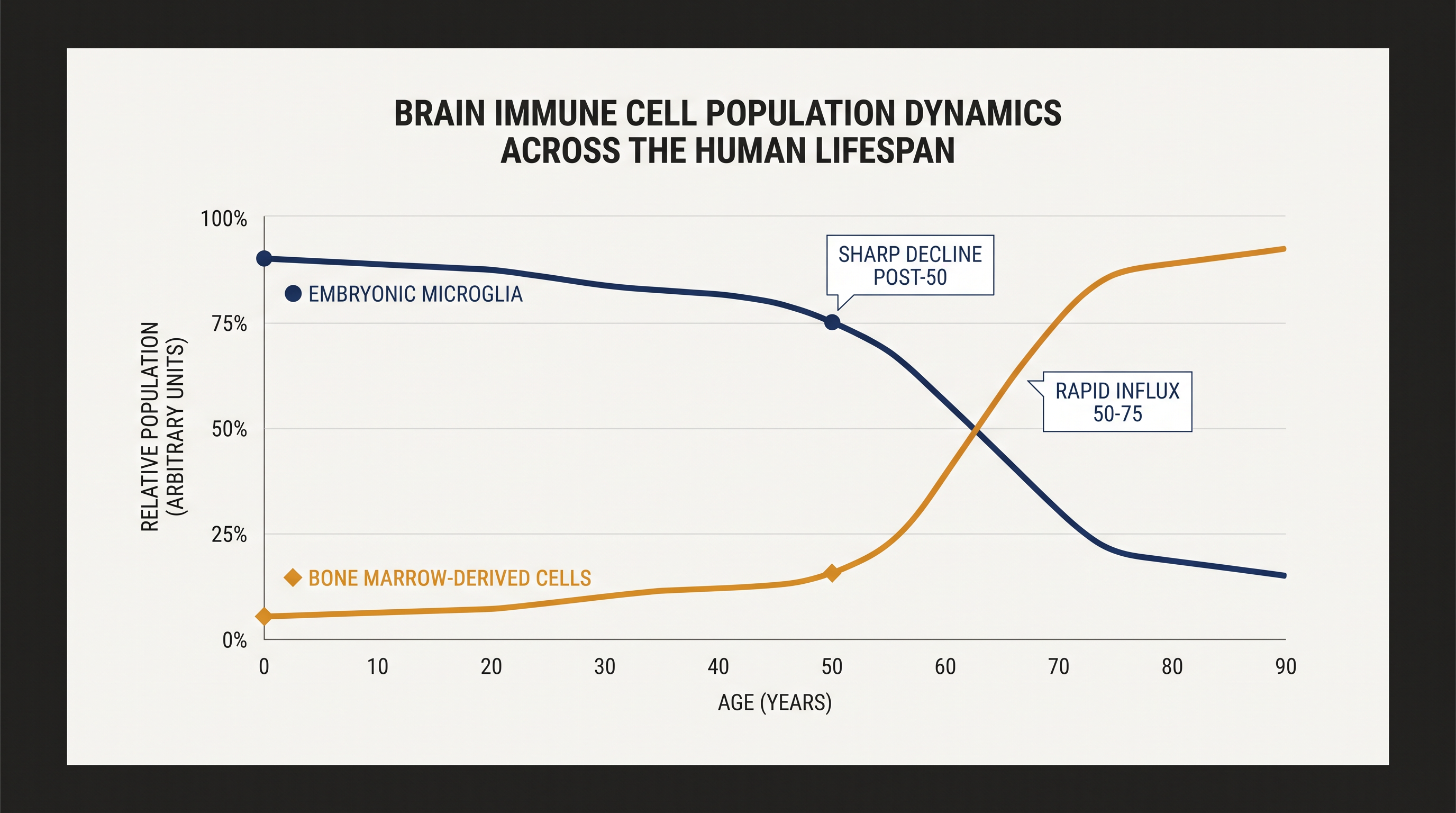
In all 20 individuals examined, marrow-derived cells had successfully crossed into the brain, taking up residence and adopting a microglia-like state.
Beyond immune turnover, single-cell profiling revealed a fundamental structural failure within the nuclei of aging brain cells, challenging previous assumptions about how gene regulation degrades over time.[1]
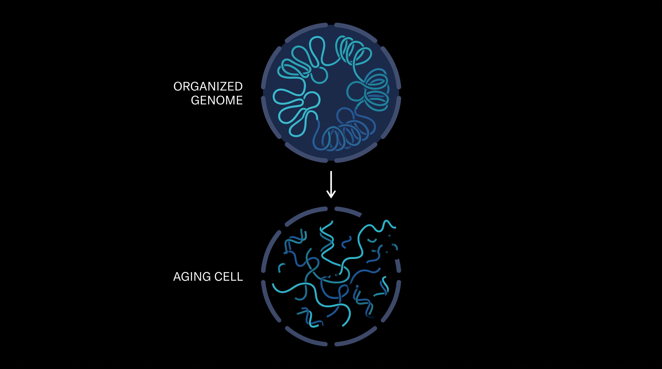
DNA is normally tightly spooled and folded into precise three-dimensional configurations, ensuring that only the correct genes are accessible and active. Across multiple brain cell types, researchers observed a global erosion of this 3D genome architecture in older tissue samples.[2]
This structural breakdown leads to widespread gene dysregulation. As the physical boundaries of the genome loosen, cells lose their specialized identities, inappropriately turning on inflammatory genes while silencing genes necessary for cellular maintenance and repair.[2][5]
The influx of bone marrow-derived cells into the brain does not happen in a vacuum, but rather coincides with a measurable decline in the specialized cell populations responsible for maintaining the blood-brain barrier.[1]
Single-cell transcriptomic clocks, which measure biological aging at the cellular level, show that the endothelial cells and pericytes lining brain capillaries undergo significant age-related transcriptomic shifts. As this barrier weakens, it provides a physical pathway for peripheral immune cells to enter the central nervous system.[2][5]
While the fact of this cellular turnover is now well-documented, a core uncertainty remains: is this immune replacement protective or destructive? Its impact on neurodegenerative diseases like Alzheimer's remains heavily contested among researchers.[6]
On one hand, the Science study data indicates that these replacement microglia-like cells exhibit elevated inflammatory signatures. This suggests that the influx of peripheral immune cells may drive the chronic neuroinflammation that is a hallmark of Alzheimer's and Parkinson's diseases.[1][2]
Conversely, the Cell study found a protective association. Patients whose blood-forming stem cells carried certain clonal hematopoiesis mutations—which were subsequently found in their brain macrophages—were statistically less likely to develop Alzheimer's disease.[3][4]
This paradox implies that while the new cells might increase baseline inflammation, they could also be more effective at clearing toxic amyloid plaques than the exhausted, aging embryonic microglia they replace. The exact conditions under which these infiltrating cells help or harm the brain remain the subject of intense ongoing study.[4][6]
Ultimately, these discoveries shift the focus of neurodegenerative research beyond the brain itself. If the brain's immune system is actively replenished by the bone marrow in later life, future therapies for Alzheimer's might target blood-forming stem cells, offering a radically new approach to treating cognitive decline.[3][6]
How we got here
Embryonic Development
Original microglia seed the brain and are sealed behind the blood-brain barrier.
Early to Mid-Adulthood
Embryonic microglia maintain brain health while the blood-brain barrier remains intact.
Age 50 to 75
The blood-brain barrier weakens, and bone marrow-derived immune cells begin infiltrating the brain.
Late Adulthood
Marrow-derived cells replace a large fraction of the original microglia, altering the brain's inflammatory profile.
Viewpoints in depth
Neuroimmunologists
Argue that the influx of peripheral immune cells drives chronic neuroinflammation and exacerbates neurodegenerative diseases.
Researchers focused on neuroinflammation point to the transcriptomic signatures of the newly arrived marrow-derived cells. These cells exhibit elevated inflammatory profiles compared to the embryonic microglia they replace. From this perspective, the age-related weakening of the blood-brain barrier is a vulnerability that allows aggressive peripheral immune cells to enter the brain, where they may inadvertently damage healthy neurons and accelerate the progression of conditions like Parkinson's and Alzheimer's.
Alzheimer's Researchers
Suggest that marrow-derived macrophages may actually protect the brain by clearing toxic amyloid plaques more effectively than exhausted original microglia.
Scientists studying the genetic drivers of Alzheimer's disease offer a counter-narrative based on large-scale sequencing cohorts. They found that patients with specific clonal hematopoiesis mutations in their blood—which subsequently migrated into their brain macrophages—had a lower risk of developing Alzheimer's. This camp argues that embryonic microglia become exhausted and senescent with age, losing their ability to clear toxic amyloid plaques. The fresh influx of marrow-derived cells, therefore, might act as a necessary reinforcement, providing the brain with capable immune scavengers just when it needs them most.
Aging Biologists
Focus on the fundamental breakdown of 3D genome architecture as the root cause of cellular confusion and aging in the brain.
For biologists studying the fundamental mechanisms of aging, the immune cell turnover is merely a symptom of a deeper structural failure. They emphasize the global erosion of 3D genome architecture observed across multiple brain cell types. When DNA unspools and loses its precise folding, cells can no longer regulate their genes properly. This camp argues that stabilizing the physical structure of the genome is the ultimate key to preventing both the degradation of the blood-brain barrier and the subsequent immune dysregulation.
What we don't know
- Whether the influx of marrow-derived immune cells can be therapeutically controlled or enhanced.
- The exact mechanisms that cause the 3D genome architecture to unspool in older brain cells.
- If preventing the breakdown of the blood-brain barrier would delay or accelerate cognitive decline.
Key terms
- Microglia
- The primary immune cells of the central nervous system, responsible for clearing cellular debris and protecting neurons.
- Somatic Mutations
- Natural, non-inherited changes in DNA that accumulate in cells over a person's lifetime.
- Transcriptomics
- The study of all RNA molecules in a cell, revealing which genes are actively turned on or off.
- Clonal Hematopoiesis
- A condition where a single mutated blood stem cell multiplies and produces a large portion of a person's blood cells.
- Blood-Brain Barrier
- A highly selective semipermeable border of cells that prevents most substances in the blood from entering the brain.
Frequently asked
Do all humans experience this immune cell turnover in the brain?
Yes, the studies found evidence of bone marrow-derived cells infiltrating the brain in all aged individuals examined.
Does this mean the brain's immune system gets stronger or weaker?
It is currently debated; the new cells may increase baseline inflammation, but they might also be better at clearing toxic proteins linked to Alzheimer's.
Can we stop the breakdown of the brain's DNA architecture?
Not yet. Understanding this 3D structural erosion is a new frontier, and researchers are currently looking for ways to stabilize the genome.
Sources
Source coverage
6 outlets
3 viewpoints surfaced





[1]ScienceNeuroimmunologists
Single-cell transcriptomic and 3D genome architecture mapping of the aging human hippocampus
Read on Science →[2]New York Genome CenterNeuroimmunologists
A single-cell study reveals major shifts in immune cells, genome organization and gene regulation in the aging human hippocampus
Read on New York Genome Center →[3]CellAlzheimer's Researchers
Clonal hematopoiesis-associated somatic mutations accumulate in microglia-like brain macrophages
Read on Cell →[4]National Institutes of HealthAlzheimer's Researchers
Somatic mutations track the infiltration of marrow-derived cells into the human brain
Read on National Institutes of Health →[5]bioRxivAging Biologists
Human cell-type-specific transcriptomic aging clocks
Read on bioRxiv →[6]Factlen Editorial Team
Synthesis by Factlen editorial team
Read on Factlen Editorial Team →

Every angle. Every day.
Get science stories with full source coverage and perspective breakdowns delivered to your inbox.