
Why the Iliotibial Band Compresses a Fat Pad Instead of Snapping Across the Knee
Anatomical research reveals that IT band syndrome is caused by the compression of a highly innervated fat pad at 30 degrees of knee flexion. This evidence debunks the long-held myth of friction and explains why stretching the band fails to resolve the pain.

In short
- Anatomical studies prove the IT band is anchored to the femur and cannot snap or rub across the knee bone, invalidating the friction theory.
- The pain of IT band syndrome comes from the band compressing a highly sensitive fat pad when the knee bends to exactly 30 degrees.
- Because the band cannot be stretched, treatment must focus on strengthening the hip abductors and increasing running cadence to reduce compressive forces.
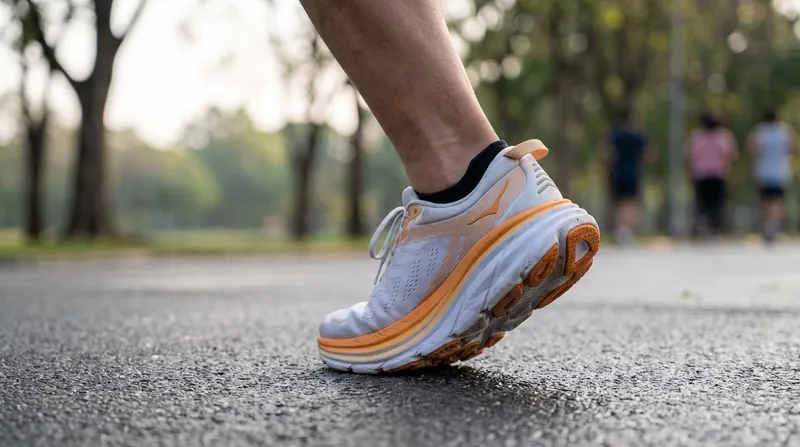
The exact moment a runner's foot strikes the ground, the knee bends to absorb the impact. As that bend reaches exactly 30 degrees of flexion, the iliotibial band pulls taut against the side of the knee. This specific angle is where the outcome of every stride is determined.[1]
At 30 degrees, the thick fascial band exerts its maximum inward pressure against the lateral femoral epicondyle, the bony prominence on the outside of the joint. If the biomechanics of the hip are aligned, this tension stabilizes the leg. If they collapse, the band crushes the tissue beneath it.[1][2]
For generations, sports medicine taught that this pain was caused by mechanical friction. The prevailing theory held that the IT band snapped back and forth across the bone like a windshield wiper, rubbing a fluid-filled sac called a bursa until it became inflamed.[5]
The Myth of Mechanical Friction
"The traditional view of ITB syndrome as a friction injury is based on a misunderstanding of the anatomy," researchers noted in a landmark 2006 anatomical study published in PubMed.[1]
Dissections of the human knee revealed that the band does not, and cannot, move back and forth across the bone. Instead of floating freely, the IT band is firmly anchored to the femur by strong fibrous bands called the linea aspera.[1]
Because it is tethered directly to the bone from the hip down to the knee, the band lacks the physical slack required to snap or roll over the epicondyle during running. If the band cannot rub, friction cannot be the source of the pain.[1]
The Compression of the Fat Pad
The 2006 anatomical review demonstrated that what actually sits beneath the IT band is not a bursa, but a highly vascularized pad of fat. When the knee bends to that critical 30-degree angle, the anchored IT band tightens and compresses this sensitive fat pad against the bone.[1][2]
"The highly innervated fat pad is the actual source of nociception in iliotibial band syndrome," the BMJ clinical review outlines.[2]
Because the tissue is packed with nerve endings and blood vessels, repeated compression during a long run triggers a severe, localized inflammatory response. The pain runners feel on the outside of the knee is the neurological response of this crushed tissue, not the chafing of a tendon.[2][3]
This compression model explains why the pain typically vanishes when the runner stops and walks. Walking requires less knee flexion at impact and generates lower ground reaction forces, keeping the knee out of the 30-degree maximum-tension window and immediately relieving the pressure on the fat pad.[3]
Why Stretching the Band Fails
Understanding that IT band syndrome is a compression injury rather than a friction problem fundamentally changes how it must be treated. For years, runners were told to stretch the band or aggressively foam-roll the outside of the thigh to lengthen the tissue and reduce the rubbing.[4][5]
The IT band is essentially a human truck tire, composed of dense, unyielding connective tissue designed to stabilize the entire weight of the body. A 2024 systematic review in Frontiers in Sports and Active Living confirmed that manual therapies cannot physically stretch or lengthen the band.[4]
"Conservative treatment strategies must focus on load management and biomechanics rather than tissue lengthening," the 2024 Frontiers review concluded.[4]
Foam rolling may temporarily desensitize the nerves in the skin, but it does not change the mechanical tension the band places on the fat pad at 30 degrees of flexion. If the band cannot be stretched, the compressive force must be reduced by changing how the leg moves.[4][6]
The Role of Hip Biomechanics
The tension on the IT band is largely dictated by the muscles at the top of the chain, specifically the gluteus medius and minimus. These two hip abductors control the inward collapse of the knee during the weight-bearing phase of the running stride.[6]
When these hip abductors fatigue during a run, the femur rotates inward and the pelvis drops on the opposite side. This structural collapse pulls the IT band tighter across the outside of the knee, drastically increasing the compressive force on the fat pad exactly when the knee hits 30 degrees.[6]
The Cleveland Clinic guidelines emphasize that strengthening the hip abductors is the most effective long-term intervention for IT band syndrome. By keeping the pelvis level and preventing the knee from diving inward, strong glutes reduce the mechanical tension on the band.[6]
Altering the Flexion Angle
Beyond building strength, runners can immediately alter the compressive forces by changing their running mechanics. Overstriding forces the knee to absorb impact at a straighter angle, often pulling it right through the 30-degree danger zone under maximum braking load.[7]
Johns Hopkins Medicine sports rehabilitation protocols recommend increasing step rate, or cadence, by 5 to 10 percent. Taking shorter, quicker steps—often targeting 165 to 175 steps per minute—brings the footstrike closer to the body's center of mass.[7]
"Increasing cadence reduces the peak hip adduction angle and the corresponding strain on the iliotibial band," clinical guidelines note.[7]
Managing the Healing Process
Because the fat pad is highly vascularized, it has an excellent capacity to heal once the excessive compression is removed. However, the PM&R Knowledge NOW clinical summary warns that returning to high-volume running too quickly will immediately re-trigger the inflammation.[3]
Rehabilitation requires a graded return to load over four to six weeks. Runners are advised to start with activities that keep the knee out of the 30-degree compression window, such as swimming or straight-leg strength work, before gradually reintroducing short, high-cadence running intervals.[3]
How we did this
- Method
- Comparing anatomical dissection data on IT band femoral anchorage against clinical treatment guidelines to derive why friction-based therapies fail while load-management succeeds.
- What we found
- The anatomical impossibility of friction directly invalidates the mechanical premise of stretching-based therapies, shifting the clinical focus entirely to compression management through hip biomechanics.
- What we worked from
- Anatomical anchorage and fat pad innervation: 30 degrees of flexion — PubMed
- Treatment efficacy of tissue lengthening: Ineffective for mechanical tension — Frontiers in Sports and Active Living
- Limits of this analysis
- This analysis relies on anatomical models and systematic reviews of conservative treatments; individual biomechanical variations may alter the exact angle of maximum compression.
Key terms
- Iliotibial band
- A thick tract of connective fascia that runs down the outside of the thigh, anchoring the hip muscles to the knee.
- Lateral femoral epicondyle
- The bony prominence on the outside of the knee joint where the IT band exerts its maximum compressive force.
- Innervated fat pad
- A layer of fat beneath the IT band that is packed with nerve endings and blood vessels, making it highly sensitive to compression.
- Knee flexion
- The anatomical term for bending the knee joint, measured in degrees from a straight leg.
Frequently asked
Will foam rolling the side of my leg cure IT band syndrome?
No. The IT band is made of dense connective tissue that cannot be lengthened by manual pressure. While foam rolling may temporarily desensitize the skin's nerve endings, it does not change the mechanical compression on the fat pad.
Should I stop running completely while the fat pad heals?
Complete rest is usually only required during the acute, highly painful phase. Once walking is pain-free, clinicians recommend a graded return to load, starting with activities that avoid the 30-degree knee flexion window, rather than resting indefinitely.
How does taking shorter steps help my knee?
Taking shorter, quicker steps (increasing cadence) brings your footstrike closer to your body's center of gravity. This changes the angle of your knee when it absorbs the impact, pulling it out of the maximum-compression zone and reducing the braking force.
Viewpoints in depth
Clinical Anatomists
Researchers who study the physical structure of the knee and its connective tissues.
Anatomists emphasize that the traditional friction model of IT band syndrome is a physical impossibility. Because the band is tethered to the femur via the linea aspera, it lacks the slack required to roll back and forth over the lateral epicondyle. Their dissections revealed that the pain originates not from a chafed bursa, but from the crushing of a highly innervated, vascularized fat pad that sits beneath the band when it pulls taut at 30 degrees of flexion.
Physical Therapists
Clinicians focused on restoring movement patterns and managing tissue load.
For physical therapists, the shift from a friction model to a compression model completely changes the rehabilitation protocol. Recognizing that the IT band is as rigid as a truck tire and cannot be lengthened by stretching or foam rolling, therapists now target the biomechanical chain. By strengthening the gluteus medius to prevent pelvic drop and increasing running cadence to alter the knee angle at impact, they reduce the compressive forces acting on the fat pad.
Traditional Sports Medicine
The historical clinical approach that dominated running injury treatment for decades.
Before the 2006 anatomical discoveries, sports medicine operated on the assumption that IT band syndrome was a friction-induced bursitis. This paradigm drove decades of treatment protocols centered on aggressively stretching the band and massaging the outer thigh to 'break up' adhesions. While these methods are now considered mechanically ineffective for lengthening the tissue, they remain deeply ingrained in running culture and are still frequently prescribed by older clinical guidelines.
- Physical Therapists
- Emphasize the biomechanical chain, prioritizing hip strength and cadence manipulation over local tissue massage.
- Clinical Anatomists
- Focus on the structural reality of the fibrous anchorage and the innervated fat pad, dismissing the friction model as physically impossible.
- Traditional Sports Medicine
- The historical perspective that relied on the bursa friction model, which drove decades of stretching and foam-rolling protocols.
Perspectives this story doesn't cover
- Footwear Biomechanists
- Recreational Running Coaches
Sources
Source coverage
8 outlets
3 viewpoints surfaced








- [1]PubMedClinical Anatomists
The functional anatomy of the iliotibial band during flexion and extension of the knee: implications for understanding iliotibial band syndrome
Read on PubMed → - [2]The BMJClinical Anatomists
Iliotibial band syndrome
Read on The BMJ → - [3]PM&R Knowledge NOWTraditional Sports Medicine
Iliotibial Band Syndrome
Read on PM&R Knowledge NOW → - [4]Frontiers in Sports and Active LivingPhysical Therapists
Effects of conservative treatment strategies for iliotibial band syndrome on pain and function in runners: a systematic review
Read on Frontiers in Sports and Active Living → - [5]NCBI BookshelfTraditional Sports Medicine
Iliotibial Band Friction Syndrome
Read on NCBI Bookshelf → - [6]Cleveland ClinicPhysical Therapists
Iliotibial Band Syndrome (ITBS)
Read on Cleveland Clinic → - [7]Johns Hopkins MedicinePhysical Therapists
Iliotibial Band Syndrome
Read on Johns Hopkins Medicine → - [8]Factlen Editorial Team
Synthesis by Factlen editorial team
Read on Factlen Editorial Team →
More in Fitness
See all →Marathon Majors
Tigst Assefa Runs 2:11:04 at Berlin Marathon, Recording Third-Fastest Time in History
5 sources
Whereabouts Rules
Olympic Silver Medalist Cyréna Samba-Mayela Provisionally Suspended for Missed Doping Tests
5 sources
World Record
Agnes Jebet Ngetich Breaks Women-Only Half Marathon World Record in Copenhagen
4 sources
Maximalist Footwear
The 35mm Threshold: How Maximalist Running Shoes Alter Foot Eversion and Hip Stability
7 sources
Comments
Every angle. Every day.
Get Fitness stories with full source coverage and perspective breakdowns, free every day.