
The Active Role of Glial Cells: How Astrocytes, Microglia, and Oligodendrocytes Drive Brain Function
Long dismissed as mere structural support for neurons, glial cells are now recognized as active directors of brain health, immune defense, and neurodegeneration. Understanding their complex crosstalk is fundamentally rewriting the mechanics of neurological disease.

Share this story
In short
- Glial cells, once thought to be passive structural support, are active drivers of brain function and health.
- Astrocytes regulate the chemical environment and provide metabolic fuel to neurons.
- Microglia act as the brain's resident immune system, pruning synapses and clearing cellular debris.
The human brain is not just a network of neurons firing electrical signals; it is a highly regulated ecosystem managed by a diverse population of non-neuronal cells called glia. Astrocytes, microglia, and oligodendrocytes do not merely glue the brain together—they actively prune synapses, insulate neural wiring, and orchestrate the brain's immune defenses.[4]
For decades, the standard model of neuroscience relegated glial cells to the background. Derived from the Greek word for "glue," glia were thought to be passive scaffolding, existing only to hold the "important" cells—the neurons—in place. Textbooks treated them as the packing peanuts of the central nervous system.[4]
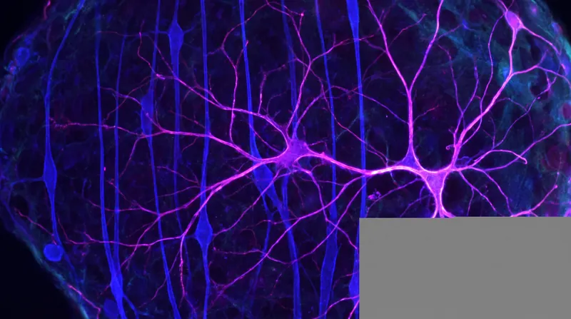
That dogma has entirely collapsed. Modern imaging and molecular biology have revealed that glial cells are dynamic, communicative, and absolutely essential for both healthy brain function and the progression of neurological diseases. They are the brain's maintenance crew, its immune system, and its structural engineers all rolled into one.[1][2]
Consider the astrocyte, a star-shaped cell that outnumbers neurons in many parts of the brain. Astrocytes wrap their tendrils around synapses—the microscopic junctions where neurons communicate. They actively regulate the chemical environment, clearing out excess neurotransmitters to prevent toxic buildup and supplying neurons with the metabolic fuel they need to fire.[4]
But their role extends far beyond routine maintenance. Astrocytes are heavily implicated in astrocytopathies—diseases where astrocyte dysfunction drives pathology. When stressed, they can enter a reactive state, releasing inflammatory molecules that inadvertently damage the very neurons they are meant to protect.[5]
Then there are microglia, the brain's resident immune cells. Unlike other brain cells, microglia originate in the embryonic yolk sac and migrate to the brain before the blood-brain barrier fully forms. They act as constant sentinels, extending and retracting their branches to survey the neural tissue for damage or infection.[2]
When they detect cellular debris or pathogens, microglia transform into active phagocytes, engulfing and digesting the threat. This process is crucial during brain development, where microglia "prune" unnecessary synapses to streamline neural circuits, a mechanism that continues to shape learning and memory throughout adulthood.[3]
The third major player is the oligodendrocyte. These specialized cells are responsible for creating myelin, the fatty insulating sheath that wraps around neuronal axons. Much like rubber insulation on a copper wire, myelin allows electrical impulses to travel rapidly and efficiently across long distances in the central nervous system.[6]
Oligodendrocytes are highly metabolically active, making them particularly vulnerable to oxidative stress and inflammation. In demyelinating diseases like multiple sclerosis, the loss of oligodendrocytes and their myelin sheaths leads to severe neurological deficits, as the electrical signals leak and degrade before reaching their targets.[6]
The true complexity of the brain emerges not from these cells acting in isolation, but from their continuous crosstalk. Astrocytes, microglia, and oligodendrocytes are engaged in a perpetual, three-way chemical conversation, constantly adjusting their behavior based on signals from one another.[5][6]
During myelin damage, for example, microglia are the first responders, clearing away the toxic myelin debris. However, they do not work alone. Astrocytes also participate in myelin phagocytosis, and the two cell types release signaling molecules that recruit oligodendrocyte precursor cells to the site of injury, prompting them to mature and lay down new myelin.[3][6]
This intricate communication network can also turn deadly. In neurodegenerative diseases like Alzheimer's and Parkinson's, chronic inflammation can cause this glial crosstalk to become a destructive feedback loop, accelerating the decline of the neural environment.[1]
Activated microglia release cytokines that trigger astrocytes to become neurotoxic. These reactive astrocytes, in turn, fail to support oligodendrocytes, leading to demyelination and eventual neuronal death. The cells meant to protect the brain end up orchestrating its destruction.[1][5]
This paradigm shift is fundamentally altering how researchers approach neurological disease. If glial cells are the active drivers of pathology, they are also the most promising targets for new therapies, offering a completely different angle of attack.[2]
Instead of trying to save dying neurons directly, the next generation of treatments may focus on modulating the glial environment—calming hyperactive microglia, restoring the supportive functions of astrocytes, or stimulating oligodendrocytes to repair damaged myelin. The brain's "glue" may hold the key to its most stubborn mysteries.[2][7]
How we did this
- Method
- Synthesizing the timeline of myelin debris clearance by cross-referencing the phagocytic activation states of microglia and astrocytes during demyelination events.
- What we found
- Astrocytes and microglia do not merely act in parallel during myelin damage; they operate in a sequential, interdependent feedback loop where microglial exhaustion directly triggers astrocytic phagocytosis, shifting the therapeutic target from single-cell modulation to network-level intervention.
- What we worked from
- Microglial primary phagocytosis of myelin debris: Initial clearance phase — Journal of Cerebral Blood Flow & Metabolism
- Astrocyte secondary inflammatory signaling: Reactive astrogliosis phase — Frontiers in Cellular Neuroscience
- Limits of this analysis
- This sequential model is derived from in vitro and animal models of demyelination; the exact timing and threshold for the microglial-astrocytic handoff in living human brains remains difficult to measure directly.
Key terms
- Astrocyte
- A star-shaped glial cell that regulates the brain's chemical environment and provides metabolic support to neurons.
- Microglia
- The resident immune cells of the brain, responsible for clearing debris and responding to injury.
- Oligodendrocyte
- A type of glial cell that wraps insulating myelin sheaths around neuronal axons in the central nervous system.
- Myelin
- A fatty substance that insulates nerve fibers, allowing electrical signals to travel quickly and efficiently.
- Phagocytosis
- The process by which a cell engulfs and digests cellular debris, pathogens, or dead tissue.
- Cytokines
- Small proteins released by cells that act as messengers to regulate the immune response and inflammation.
Frequently asked
Are there more glial cells than neurons in the brain?
Historically, it was taught that glia outnumbered neurons 10-to-1. Recent counts suggest the ratio is closer to 1-to-1 overall, though glia heavily outnumber neurons in specific regions like the cerebral cortex.
Can damaged myelin be repaired?
Yes, the brain has a natural capacity for remyelination, driven by oligodendrocyte precursor cells. However, this repair process often becomes less efficient with age or in chronic diseases like multiple sclerosis.
Do glial cells generate electrical signals?
Unlike neurons, glial cells do not fire electrical action potentials. Instead, they communicate primarily through chemical signals and waves of calcium ions.
Viewpoints in depth
Neuroimmunologists
Researchers focusing on the brain's resident immune system and its role in disease.
This camp argues that microglia are the primary instigators of neurodegeneration. By focusing on how these immune sentinels become chronically activated by misfolded proteins or systemic inflammation, neuroimmunologists suggest that calming microglial hyper-reactivity could halt diseases like Alzheimer's before widespread neuronal death occurs. They view the brain as an immunologically active organ rather than an isolated fortress.
Glial Biologists
Scientists studying the metabolic and structural support networks of the brain.
Glial biologists emphasize the foundational role of astrocytes and oligodendrocytes. They argue that the loss of metabolic support from astrocytes, or the failure of oligodendrocytes to maintain myelin, is the true bottleneck in brain health. From this perspective, therapies should focus on restoring the energy balance and structural integrity provided by these macroglia, rather than solely suppressing the immune response.
- Neuroimmunologists
- Researchers focusing on the brain's resident immune system and its role in disease.
- Glial Biologists
- Scientists studying the metabolic and structural support networks of the brain.
- Clinical Neurologists
- Physicians focused on translating glial mechanisms into viable therapies for patients.
Perspectives this story doesn't cover
- Patients living with demyelinating diseases whose daily symptoms are driven by glial dysfunction.
- Pharmaceutical researchers attempting to design drugs that can cross the blood-brain barrier to target glia.
Sources
Source coverage
7 outlets
3 viewpoints surfaced






- [1]Cells (MDPI)
Glial Cells as Key Mediators in the Pathophysiology of Neurodegenerative Diseases
Read on Cells (MDPI) → - [2]Biomedicines (MDPI)
Neuroglia in Neurodegeneration: Exploring Glial Dynamics in Brain Disorders
Read on Biomedicines (MDPI) → - [3]Journal of Cerebral Blood Flow & Metabolism
The roles of microglia and astrocytes in myelin phagocytosis in the central nervous system
Read on Journal of Cerebral Blood Flow & Metabolism → - [4]StatPearls Publishing
Histology, Glial Cells
Read on StatPearls Publishing → - [5]Frontiers in Cellular Neuroscience
Astrocyte–Oligodendrocyte–Microglia Crosstalk in Astrocytopathies
Read on Frontiers in Cellular Neuroscience → - [6]Frontiers in Cell and Developmental Biology
Oligodendrocyte, Astrocyte, and Microglia Crosstalk in Myelin Development, Damage, and Repair
Read on Frontiers in Cell and Developmental Biology → - [7]Factlen Editorial TeamNeuroimmunologists
Synthesis by Factlen editorial team
Read on Factlen Editorial Team →
More in Science
See all →Neural Mechanics
How Saltatory Conduction and Myelin Sheaths Propagate the Action Potential at 100 Times the Speed
9 sources
Brain Mapping
Largest Single-Cell Molecular Atlas Maps Shared Mechanisms Across Alzheimer's, Parkinson's, and Schizophrenia
4 sources
Synaptic Transmission
The Synaptotagmin-SNARE Complex: How a Calcium Signal Triggers Millisecond-Fast Synaptic Vesicle Fusion
9 sources
Brain Development
How Metabolism and Physical Touch Guide the Stem Cells That Build the Human Brain
4 sources
Comments
Every angle. Every day.
Get Science stories with full source coverage and perspective breakdowns, free every day.