
How Neural Gliding Mechanically Frees Entrapped Nerves Without Stretching Tissue
Peripheral nerves lose their blood supply when stretched beyond 15 percent of their resting length. A technique called neural gliding restores mobility by sliding the nerve through its sheath rather than pulling it taut.
By Jun Zhao

Share this story
The central nervous system operates as the ultimate governor of human mobility, determining exactly how far a limb can extend and when that range can safely increase. When a peripheral nerve becomes entrapped or irritated, the system's immediate response is to lock down the surrounding tissue. This creates a sensation of profound tightness that athletes often mistake for a shortened muscle. But while a muscle belly can be forced to lengthen through sustained static stretching, applying that same mechanical load to a compromised nerve reliably makes the entrapment worse.[2][4]
The distinction comes down to tissue architecture. Skeletal muscle is highly elastic, designed to stretch up to 30 percent beyond its resting length to absorb and generate force. Peripheral nerves, by contrast, are viscoelastic cables that prioritize signal transmission over structural elasticity. They do not stretch; they slide. When an athlete attempts to "stretch out" a sharp, radiating pain in their hamstring or calf, they are often pulling a tethered nerve against a mechanical restriction, triggering a cascade of protective inflammation.[1][2]
"Using a stereo-imaging camera system provides a non-invasive method for capturing three-dimensional displacements of peripheral nerves when stretched," writes Virginia Orozco, lead author of a 2024 methodological study published in the Journal of Visualized Experiments. Her team's work highlights how nerves respond to tension in real time. At roughly 8 percent elongation, venous blood flow inside a peripheral nerve begins to slow. By 15 percent elongation, arterial flow stops entirely, inducing localized ischemia.[3]
This ischemic threshold explains why traditional flexibility protocols fail for nerve pain. Holding a static stretch for 30 to 60 seconds starves the entrapped nerve of oxygen. The surrounding fascia thickens in response to the perceived trauma, tightening the mechanical bottleneck. What feels like a stubborn muscle knot is actually a neurological safeguard, preventing the athlete from severing their own wiring.[1][4]
The clinical alternative is neurodynamics, commonly known as nerve flossing or neural gliding. Instead of anchoring both ends of the tissue and pulling the middle taut, flossing involves moving the body to pull the nerve from one direction while simultaneously releasing tension from the other. The movement mimics a piece of dental floss sliding back and forth between two teeth, freeing the nerve from adhesions without ever subjecting it to a sustained stretch.[2]
"Understanding the biomechanical response of peripheral nerves to stretch is critical to the understanding of their response to different loading conditions and thus, to optimizing treatment strategies," notes Anita Singh of Temple University's Bioengineering Department, a co-author on the 2024 strain study. By keeping the total elongation below the 8 percent ischemic threshold, flossing restores the nerve's natural glide path while maintaining continuous blood flow.[3]
Differentiating between muscle soreness and nerve irritation dictates the recovery protocol. Delayed onset muscle soreness (DOMS) typically peaks between 48 and 72 hours after a heavy training load. It presents as a dull, widespread ache that improves with light movement and static stretching. Nerve pain, however, is sharp, electrical, and often radiates down the limb. It does not fade on a predictable three-day timeline, and it sharply worsens when the limb is held in a stretched position.[2][5]
The dosage for neural gliding differs entirely from muscle hypertrophy or flexibility training. Because nerves are highly sensitive to mechanical friction, physical therapists typically prescribe low-volume interventions—often just 5 to 10 repetitions per session, performed two to three times a day. Pushing through pain or accumulating high rep counts inflames the nerve sheath, setting the recovery process back by several days.[2]
For the median nerve, which runs down the inside of the arm and is frequently implicated in carpal tunnel syndrome, the glide involves extending the arm and wrist while tilting the head toward the working shoulder. As the wrist extends (pulling the nerve distally), the neck tilts (creating slack proximally). The sequence is then reversed, sliding the nerve smoothly through the carpal tunnel without stretching it.[1][2]
Similar protocols exist for the sciatic nerve in the lower body and the ulnar nerve in the arm. In high-load movements like the Farmer's Walk, where athletes carry heavy weights over distance, the cervical and brachial nerves are subjected to immense traction. If an athlete develops radiating pain down their arm after a heavy carrying session, aggressive neck stretching is the exact wrong intervention. A targeted ulnar or radial nerve glide resolves the entrapment by restoring mobility, not length.[1][6]
The shift from stretching to flossing represents a broader evolution in sports medicine. Mobility is no longer viewed purely as a mechanical property of muscle and fascia, but as a neurological permission slip granted by the central nervous system. By respecting the ischemic limits of peripheral nerves, athletes can clear stubborn movement restrictions that traditional flexibility work could never reach.
Viewpoints in depth
Clinical Biomechanists
Researchers focused on the mechanical limits and ischemic thresholds of peripheral nerve tissue.
This camp emphasizes that nerves are viscoelastic structures designed for signal transmission, not mechanical load-bearing. By measuring in-situ strain during stretching, biomechanists have demonstrated that applying static tension to a nerve rapidly cuts off its blood supply. They argue that understanding these precise failure points—such as the 15 percent elongation threshold for arterial ischemia—is essential for developing safe rehabilitation protocols that do not inadvertently damage the tissue they intend to heal.
Physical Therapists
Rehabilitation professionals who utilize neurodynamics to treat entrapment syndromes.
Physical therapists view mobility restrictions through a neurological lens, recognizing that what feels like a "tight muscle" is often the central nervous system locking down a joint to protect an irritated nerve. They advocate for nerve flossing as a primary intervention for conditions like sciatica and carpal tunnel syndrome. Their protocols prioritize low-repetition, pain-free movements that restore the nerve's natural glide path, explicitly warning patients against pushing into the stretch or holding tension for prolonged periods.
Strength & Conditioning Coaches
Training professionals focused on managing athlete recovery and differentiating types of tissue stress.
For coaches managing high training loads, the priority is distinguishing between normal muscular adaptation (DOMS) and neurological warning signs. They monitor recovery timelines closely, knowing that while muscle soreness peaks at 48 to 72 hours and responds well to active recovery, sharp or radiating pain indicates nerve involvement. This camp integrates targeted nerve glides into mobility warm-ups, particularly for athletes performing heavy loaded carries or overhead lifts that place immense traction on the cervical and brachial plexus.
What we don’t know
- Whether prophylactic nerve flossing prevents entrapment syndromes in healthy athletes, or if it is only effective as a rehabilitative tool once irritation has occurred.
- The exact variation in ischemic thresholds across different peripheral nerves, as most in-situ strain data is derived from the sciatic and sural nerves.
- How long-term neurodynamic training alters the structural thickness of the myelin sheath over years of practice.
- Clinical Biomechanists
- Researchers focused on the mechanical limits and ischemic thresholds of peripheral nerve tissue.
- Physical Therapists
- Rehabilitation professionals who utilize neurodynamics to treat entrapment syndromes.
- Strength & Conditioning Coaches
- Training professionals focused on managing athlete recovery and differentiating types of tissue stress.
Sources
Source coverage
6 outlets
3 viewpoints surfaced





- [1]Factlen Editorial TeamPhysical Therapists
Synthesis by Factlen editorial team
Read on Factlen Editorial Team → - [2]Women's HealthPhysical Therapists
Nerve Flossing May Help Ease Pain—Here's How It Works and What Experts Want You to Know
Read on Women's Health → - [3]Journal of Visualized ExperimentsClinical Biomechanists
Direct Linear Transformation for the Measurement of In-Situ Peripheral Nerve Strain During Stretching
Read on Journal of Visualized Experiments → - [4]Women's HealthPhysical Therapists
6 Signs You’re Running Too Much, According To A Physical Therapist
Read on Women's Health → - [5]Runner's WorldStrength & Conditioning Coaches
This 3-Day Soreness Checklist Can Reveal If You Nailed Your Workout—or Pushed Too Hard
Read on Runner's World → - [6]Men's HealthStrength & Conditioning Coaches
Build Real Total-Body Strength With the Farmer's Walk
Read on Men's Health →
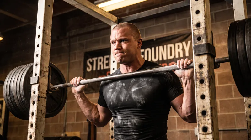
More in Fitness
See all →Wearable Tech
Amazfit Firmware Update Adds FTMS Smart Trainer Support and On-Watch HYROX Analysis
4 sources
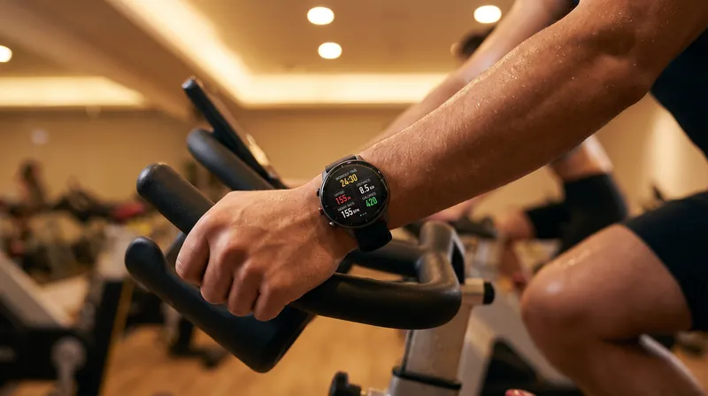
Stretch-Shortening Cycle
Why Tendons Only Stiffen When Ground Contact Drops Below 250 Milliseconds
4 sources

Combat Readiness
Pentagon Codifies Sex-Neutral Fitness Standards Across All Military Branches
4 sources
Metabolic Science
Pterostilbene's PPAR-Delta Stabilization: How a Blueberry Compound Forces Skeletal Muscle to Break Down Fat
6 sources

Comments
Every angle. Every day.
Get Fitness stories with full source coverage and perspective breakdowns, free every day.